. 2010 Jun;16(6):255-264.
doi: 10.1016/j.mpdhp.2010.03.008. Epub 2010 Apr 21.

Inflammatory diseases of the nasal cavities and paranasal sinuses

Affiliations

Inflammatory diseases of the nasal cavities and paranasal sinuses

Tim Helliwell. Diagn Histopathol (Oxf). 2010 Jun.

Abstract

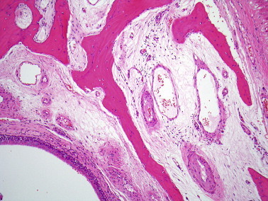
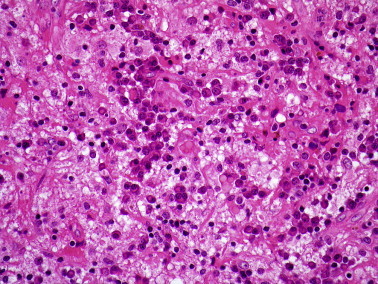
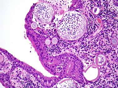
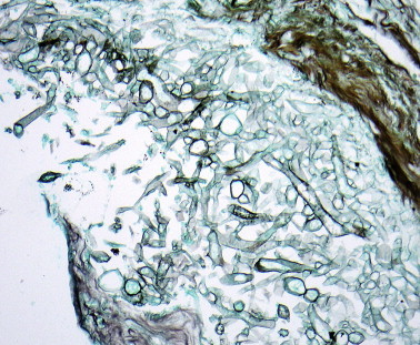
Inflammatory diseases of the nose and paranasal sinus are commonly encountered in diagnostic histopathology. This review describes the possible manifestations of the common diseases as well as highlighting some of the uncommon causes of sinonasal inflammation which may have importance for treatment and prognosis. The diagnosis of fungal sinusitis is primarily histological. It is important to distinguish between invasive and non-invasive fungal sinusitis, the latter including allergic fungal sinusitis characterized by 'allergic mucin' and scanty fungal hyphae. Nasal eosinophilia is a feature of both allergic and non-allergic rhinosinusitis and a wide range of secondary changes in inflammatory polyps may lead to diagnostic confusion. Nasal biopsies are often taken from perforations or inflammatory masses to confirm or exclude granulomatous diseases. There is a broad differential diagnosis for granulomatous sinonasal disease and pathologists should appreciate the diagnostic histological and clinical features of these conditions.

Keywords: chronic inflammation; diagnosis; fungal diseases; granulomatous inflammation; nose and paranasal sinuses; pathology.


Figures

Figure 1
Figure 1
Normal nasal mucosa is covered by respiratory epithelium and contains many vessels.
Figure 2
Figure 2
Normal sinus mucosa is thinner and closely apposed to bone.
Figure 3
Figure 3
Rhinoscleroma showing large vacuolated macrophages, plasma cells and lymphocytes.
Figure 4
Figure 4
Rhinosporidiosis with large sporangia containing endospores in the superficial mucosal tissue.
Figure 5
Figure 5
Broad irregular hyphae of Mucor sp. (Grocott silver stain).
Figure 6
Figure 6
Dichotomously branching septate hyphae of Aspergillus sp.
Figure 7
Figure 7
Laminated masses of fungal hyphae in a sinus fungal ball.
Figure 8
Figure 8
‘Allergic mucin’ composed of inspissated eosinophilic mucin containing masses of degenerate eosinophils and epithelial cells.
Figure 9
Figure 9
Inflammatory nasal polyp formed by oedematous, pale brown mucosal tissue.
Figure 10
Figure 10
Immature squamous metaplasia on the surface of an inflammatory polyp.
Figure 11
Figure 11
Inflammatory nasal polyp covered by respiratory epithelium with basement membrane thickening and an eosinophil-rich inflammatory infiltrate.
Figure 12
Figure 12
Inflammatory polyp showing extensive fibrinous exudation that superficially resembles amyloid.
Figure 13
Figure 13
Inflammatory polyp with marked glandular hyperplasia.
Figure 14
Figure 14
Cholesterol granuloma in an inflammatory polyp associated with haemorrhage.
Figure 15
Figure 15
Nasopharyngeal angiofibroma is formed from plump spindle cells which surround thick-walled vessels.
Figure 16
Figure 16
Seromucinous hamartoma showing small glands that lack the normal lobularity of mucosal glands.
Figure 17
Figure 17
Foreign material in the inflammatory exudate from a patient with septal perforation associated cocaine inhalation.
Figure 18
Figure 18
Granulomatous inflammation due to sarcoidosis has well defined epithelioid cell granulomas.
Figure 19
Figure 19
Granulomatous inflammation in Wegener's granulomatosis shows poorly defined epithelial cell granulomas, multinucleate cells and lymphocytes.
Figure 20
Figure 20
Inflammatory destruction of cartilage in relapsing polychondritis.
Figure 21
Figure 21
a Eosinophilic angiocentric fibrosis showing nodular fibrosis around vessels. b Very few eosinophils are present at this stage of the disease.

Similar articles

Cited by

References

    1. Kim Y.M., Kim A.Y., Park Y.H., Kim D.H., Rha K.S. Eight cases of nasal tuberculosis. Otolaryngol Head Neck Surg. 2007;137:500–504. - PubMed
    1. Melo Naves M., Gomes Patrocinio L., Patrocinio J.A. Contribution of nasal biopsy to leprosy diagnosis. Am J Rhinol Allergy. 2009;23:177–180. - PubMed
    1. de Pontual L., Ovetchkine P., Rodriguez D. Rhinoscleroma: a French national retrospective study of epidemiological and clinical features. Clin Infect Dis. 2008;47:1396–1402. - PubMed
    1. Hart C.A., Rao S.K. Rhinoscleroma. J Med Microbiol. 2000;49:395–396. - PubMed
    1. Fredricks D.N., Jolley J.A., Lepp P.W., Kosek J.C., Relman D.A. Rhinosporidium seeberi: a human pathogen from a novel group of aquatic protistan parasites. Emerg Infect Dis. 2000;6:273–282. - PMC - PubMed

LinkOut - more resources