. 2020 Jun 3;29(9):1520-1536.
doi: 10.1093/hmg/ddaa075.

A rare genomic duplication in 2p14 underlies autosomal dominant hearing loss DFNA58

Affiliations

A rare genomic duplication in 2p14 underlies autosomal dominant hearing loss DFNA58

Karina Lezirovitz et al. Hum Mol Genet. .

Abstract

Here we define a ~200 Kb genomic duplication in 2p14 as the genetic signature that segregates with postlingual progressive sensorineural autosomal dominant hearing loss (HL) in 20 affected individuals from the DFNA58 family, first reported in 2009. The duplication includes two entire genes, PLEK and CNRIP1, and the first exon of PPP3R1 (protein coding), in addition to four uncharacterized long non-coding (lnc) RNA genes and part of a novel protein-coding gene. Quantitative analysis of mRNA expression in blood samples revealed selective overexpression of CNRIP1 and of two lncRNA genes (LOC107985892 and LOC102724389) in all affected members tested, but not in unaffected ones. Qualitative analysis of mRNA expression identified also fusion transcripts involving parts of PPP3R1, CNRIP1 and an intergenic region between PLEK and CNRIP1, in the blood of all carriers of the duplication, but were heterogeneous in nature. By in situ hybridization and immunofluorescence, we showed that Cnrip1, Plek and Ppp3r1 genes are all expressed in the adult mouse cochlea including the spiral ganglion neurons, suggesting changes in expression levels of these genes in the hearing organ could underlie the DFNA58 form of deafness. Our study highlights the value of studying rare genomic events leading to HL, such as copy number variations. Further studies will be required to determine which of these genes, either coding proteins or non-coding RNAs, is or are responsible for DFNA58 HL.


Figures

Figure 1
Figure 1
Pedigree showing the segregation of the DFNA58 duplication (− =non-duplicated; D = duplicated) and flanking microsatellite markers haplotype. Expression data from CNRIP1, LOC102724389 and LOC107985892 (mRNA) are also shown in the box below the haplotypes of each subject: +++ means RNA overexpression in RT-qPCR; −— means RNA normal expression in RT-qPCR.
Figure 2
Figure 2
(A) Age distribution of onset of 17 patients with the DFNA58 HL. (B) 25 audiometric profiles of 17 affected patients divided in in four different age at exam groups. Subject IV:2 had a cholesteatoma in her right ear, in addition to the bilateral postlingual progressive sensorineural HL, thus explaining why her HL is not symmetrical, being profound in the right ear and moderate in the left ear. (C) Distribution of average thresholds of six affected individuals according to their age at examination showing fit to a linear regression, only when excluding the subject IV:6 with childhood onset.
Figure 3
Figure 3
(A) Multipoint LOD scores calculated with Morgan software for markers in the 2p12-p21 chromosomal region using a penetrance estimate of 96%. (B) Sketch of the duplication showing the location of MLPA probes, as well as extension of the duplication according to the different methods and combining all [NC_000002.12:g.(68 247 572_68 248 077)_(68 449 525_68 452 166)dup]. The arrows indicate transcription orientation. The exons of each gene producing the longest mRNA and with RefSeq entry are represented, GRCh38 as the reference genome assembly. Both breakpoints (BK regions) locate within repetitive elements. Circled Genes are observed overexpression in the blood of duplication carriers are indicated by a circle.
Figure 4
Figure 4
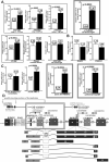
(AC) RT-qPCR results showing mRNA blood levels in duplication carriers (mean of at least 10 subjects, indicated in black) and non-carriers (mean of at least eight subjects, indicated in white) of genes involved in the duplication or close to its probable insertion position: (A) Genes involved in the duplication: PPP3R1 (exon 1), CNRIP1 (all transcripts), PLEK (protein coding transcript and retained intron transcript) and PPP3R1(overall expression of all transcripts). (B) mRNA levels of PNO1 and WDR92, two neighbor genes of the possible duplication insertion position. (C) mRNA levels of uncharacterized lncRNA genes contained inside the duplication segment: LOC101927723, AC017083.1, LOC102724389 and LOC107985892. Graphs showing genes that are mildly overexpressed (1.2–1.8X) were indicated by a single line border: PLEK (protein coding transcript), PPP3R1 (exon 1 of all three transcripts) and AC017083.1 (lncRNA gene). Graphs showing genes that were highly overexpressed (4-27X) comparing the means of carriers X non-carriers were indicated by double line border: CNRIP1, LOC102724389 and LOC107985892. (D) Possible insertion positions of the duplicated segment (?). The size of the duplicated segment is indicated by double line border. Depending on the insertion position, the new gene AC017083.3 may or may not be interrupted. Examples of the low-abundant fusion CNRIP1-PPP3R1 transcripts found in the duplication carriers are shown below.
Figure 5
Figure 5
ISH in sagittal sections of P7, P21 and P35 mouse cochlea showing the expression of the candidate gene Cnrip1. AS, antisense probe; S, sense probe (negative control).White scale: 200 μm; Gray scale: 50 μm. White arrowhead: SGN; yellow dotted contour: inner and outer hair cells. The hair-cell specific marker Loxhd1, used as control, is in Supplementary Figure S2.
Figure 6
Figure 6
ISH in sagittal sections of P7, P21 and P35 mouse cochlea showing the expression of the candidate gene Plek.AS, antisense probe; S, sense probe (negative control). White scale: 200 μm; Gray scale: 50 μm. White arrowhead: SGN; yellow dotted contour: inner and outer hair cells. The hair-cell specific marker Loxhd1, used as control, is in Supplementary Figure S2.
Figure 7
Figure 7
ISH in sagittal sections of P7, P21 and P35 mouse cochlea showing the expression of the candidate gene Ppp3r1. AS, antisense probe; S, sense probe (negative control). White scale: 200 μm; Gray scale: 50 μm. White arrowhead: SGN; white dotted contour: pillar cells; yellow dotted contour: inner and outer hair cells. The hair-cell specific marker Loxhd1, used as control, is in Supplementary Figure S2.
Figure 8
Figure 8
CNRIP1 immunofluorescence in sagittal sections of young adult murine cochleae. (A) Relatively intense CNRIP1 staining (green) of SGNs located at the periphery of the spiral ganglion at P21. Some nuclei were stained within the spiral ligament and stria vascularis. Actin stained using fluorescently tagged phalloidin (magenta). (B) At P35, neurons most strongly expressing CNRIP1 double labeled using anti-peripherin (magenta), a specific marker of type II SGNs. Anti-CNRIP1 immunofluorescence also detected in peripheral nerve fibers leaving the ganglion, toward the auditory brainstem. Scale bars = 50 μm.
Figure 9
Figure 9
PLEK immunofluorescence in sagittal sections of young adult murine cochleae. (A) In the basal turn at P35, PLEK immunofluorescence (green) evident within SGN cell bodies and fibrocytes on the perilymphatic face of spiral limbus. Actin stained using fluorescently tagged phalloidin (magenta). (B) Detail of the spiral ganglion in the basal cochlear region. Scale bars = 50 μm.

References

    1. Hilgert N., Smith R.J. and Van Camp G. (2009) Forty-six genes causing nonsyndromic hearing impairment: which ones should be analyzed in DNA diagnostics? Mutat. Res., 681, 189–196. - PMC - PubMed
    1. NIDCD Epidemiology and Statistics Program Age at which hearing loss begins. Source: National Health Interview Survey, 2007https://www.nidcd.nih.gov/health/statistics/age-which-hearing-loss-begins(updated in November 2012).
    1. Op de Beeck K., Schacht J. and Van Camp G. (2011) Apoptosis in acquired and genetic hearing impairment: the programmed death of the hair cell. Hear. Res., 281, 18–27. - PMC - PubMed
    1. Bowl M.R. and Brown S.D.M. (2018) Genetic landscape of auditory dysfunction. Hum. Mol. Genet., 27, R130–R135. - PubMed
    1. Yan D., Zhu Y., Walsh T., Xie D., Yuan H., Sirmaci A., Fujikawa T., Wong A.C., Loh T.L., Du L. et al. (2013) Mutation of the ATP-gated P2X(2) receptor leads to progressive hearing loss and increased susceptibility to noise. Proc. Natl. Acad. Sci. USA., 110, 2228–2233. - PMC - PubMed

Publication types

MeSH terms