

Total reflection X-ray fluorescence spectrometry for trace determination of iron and some additional elements in biological samples
- PMID: 32337622
- PMCID: PMC7442763
- DOI: 10.1007/s00216-020-02614-8
Total reflection X-ray fluorescence spectrometry for trace determination of iron and some additional elements in biological samples
Abstract
Trace elements are essential for life and their concentration in cells and tissues must be tightly maintained and controlled to avoid pathological conditions. Established methods to measure the concentration of trace elements in biological matrices often provide only single element information, are time-consuming, and require special sample preparation. Therefore, the development of straightforward and rapid analytical methods for enhanced, multi-trace element determination in biological samples is an important and raising field of trace element analysis. Herein, we report on the development and validation of a reliable method based on total reflection X-ray fluorescence (TXRF) analysis to precisely quantify iron and other trace metals in a variety of biological samples, such as the liver, parenchymal and non-parenchymal liver cells, and bone marrow-derived macrophages. We show that TXRF allows fast and simple one-point calibration by addition of an internal standard and has the potential of multi-element analysis in minute sample amounts. The method was validated for iron by recovery experiments in homogenates in a wide concentration range from 1 to 1600 μg/L applying well-established graphite furnace atomic absorption spectrometry (GFAAS) as a reference method. The recovery rate of 99.93 ± 0.14% reveals the absence of systematic errors. Furthermore, the standard reference material "bovine liver" (SRM 1577c, NIST) was investigated in order to validate the method for further biometals. Quantitative recoveries (92-106%) of copper, iron, zinc, and manganese prove the suitability of the developed method. The limits of detection for the minute sample amounts are in the low picogram range. Graphical abstract.
Keywords: Biometal trace analysis; Bone marrow–derived macrophages; Iron trace analysis; Liver cells; Liver tissues; Total reflection X-ray fluorescence spectrometry.
Conflict of interest statement
The authors declare that they have no conflicts of interest.
Figures


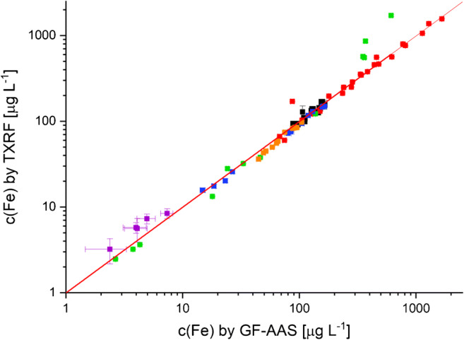
 , HSC;
  , HC;
, HC;  , LSEC;
  , KC), macrophages (
 , BMDM), and liver tissues (
 ) by TXRF using GFAAS as a standard reference method (recovery function: y = (0.9993 ± 0.0014)x − (2.8456 ± 0.3332); error bars represent uncertainties as derived from calibration prognosis interval for GFAAS measurement and from errors calculated from the spectra for TXRF measurements)
References
-
- Carvalho ML, Magalhães T, Becker M, von Bohlen A. Trace elements in human cancerous and healthy tissues: a comparative study by EDXRF, TXRF, synchrotron radiation and PIXE. Spectrochim Acta Part B At Spectrosc. 2007;62:1004–1011. doi: 10.1016/j.sab.2007.03.030. - DOI
MeSH terms
Substances
Grants and funding
LinkOut - more resources
Full Text Sources
Medical
