

Bacterial rhomboid proteases mediate quality control of orphan membrane proteins
- PMID: 32337752
- PMCID: PMC7232013
- DOI: 10.15252/embj.2019102922
Bacterial rhomboid proteases mediate quality control of orphan membrane proteins
Abstract
Although multiprotein membrane complexes play crucial roles in bacterial physiology and virulence, the mechanisms governing their quality control remain incompletely understood. In particular, it is not known how unincorporated, orphan components of protein complexes are recognised and eliminated from membranes. Rhomboids, the most widespread and largest superfamily of intramembrane proteases, are known to play key roles in eukaryotes. In contrast, the function of prokaryotic rhomboids has remained enigmatic. Here, we show that the Shigella sonnei rhomboid proteases GlpG and the newly identified Rhom7 are involved in membrane protein quality control by specifically targeting components of respiratory complexes, with the metastable transmembrane domains (TMDs) of rhomboid substrates protected when they are incorporated into a functional complex. Initial cleavage by GlpG or Rhom7 allows subsequent degradation of the orphan substrate. Given the occurrence of this strategy in an evolutionary ancient organism and the presence of rhomboids in all domains of life, it is likely that this form of quality control also mediates critical events in eukaryotes and protects cells from the damaging effects of orphan proteins.
Keywords: Shigella; intramembrane proteolysis; membrane protein complexes; quality control; rhomboid.
© 2020 The Authors. Published under the terms of the CC BY 4.0 license.
Conflict of interest statement
The authors declare that they have no conflict of interest.
Figures
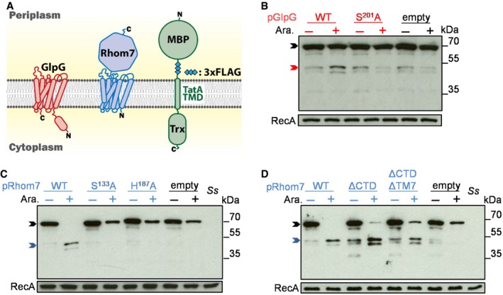
Topology of GlpG, Rhom7 and the artificial substrate with a maltose‐binding protein (MBP) domain, triple‐FLAG tag (3xFLAG), the TMD of P. stuartii TatA and a thioredoxin domain (Trx).
Western blot analysis (probing with an anti‐FLAG mAb) to detect cleavage of the artificial substrate by wild‐type (WT)/inactive (S201A) GlpG encoded on pBAD33 with/without arabinose (Ara.).
Western blot analysis to detect cleavage of the artificial substrate by wild‐type (WT)/modified (S133A or H187A) Rhom7 encoded on pBAD33 with/without arabinose (Ara.).
Activity of Rhom7 with/without its 7th TMD and/or C‐terminal domain.
- A, B
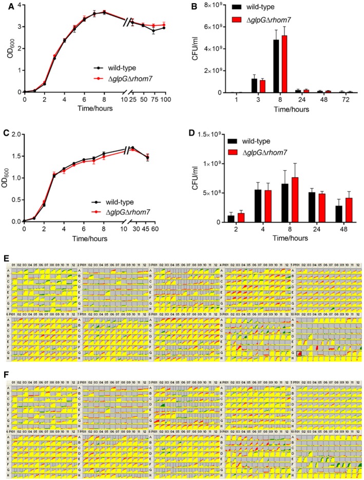
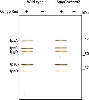
Wild‐type S. sonnei or S. sonnei ΔglpGΔrhom7 were grown in liquid LB in the presence of oxygen at 37°C with shaking at 180 rpm. Growth was assessed by measuring the optical density of cultures at 600 nm (OD600) (A) and colony‐forming units of bacteria (CFU) recovered from the same samples (B).
- C, D
Bacteria were grown anaerobically in LB supplemented with 10 mM KNO3 with shaking at 180 rpm. Growth was assessed by measuring the OD600 as above (C) and CFU of bacteria recovered from the same cultures (D).
- E, F
Comparisons of cellular respiration (NADH production) of S. sonnei ΔglpGΔrhom7 (green) to wild‐type S. sonnei (red) (E), or of S. sonnei ΔglpGΔrhom7 + glpG (green) to wild‐type S. sonnei (red) (F) in conditions provided by PM1‐10 of the Biolog MicroArrays. Areas of overlap are coloured yellow, while differences are shown as patches of red or green. Wild‐type S. sonnei seemed to respire better than S. sonnei ΔglpGΔrhom7 + glpG in pH 9.5 + anthranilic acid (black box in E). However, this result was not reproducible.
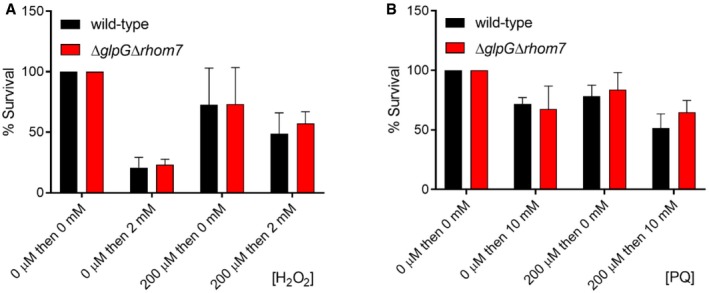
Resistance of wild‐type S. sonnei or S.sonnei ΔglpGΔrhom7 to H2O2. S. sonnei was grown in LB to an OD600 of ≈ 0.5 and then pre‐incubated in 0 or 200 μM H2O2 for 20 min, before incubation in 0 or 2 mM H2O2 for 15 min. Bacterial survival was measured by plating samples to solid media. The percentage survival was calculated as the ratio of CFU recovered from H2O2‐exposed bacteria compared to bacteria not exposed to H2O2.
Resistance of wild‐type S. sonnei or S. sonnei ΔglpGΔrhom7 to paraquat (PQ). S. sonnei was grown as above and then pre‐incubated in 0 or 200 μM PQ for 15 min before incubation in 0 or 10 mM PQ for 45 min; samples were then plated to solid media to measure bacteria survival. The percentage survival was calculated as the ratio of CFU recovered from PQ‐treated bacteria to those that were untreated.
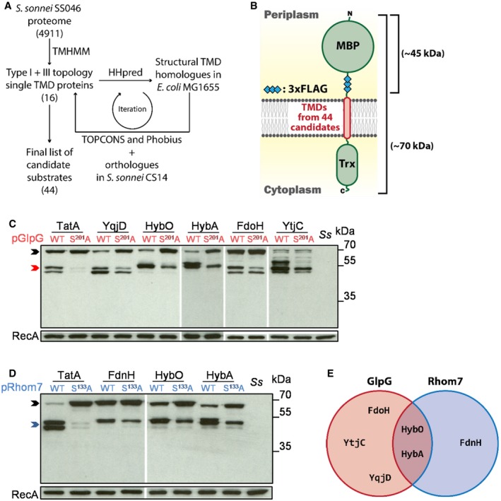
Workflow of the bioinformatic identification of candidate rhomboid substrates.
A library of artificial substrates harbouring TMDs from 44 candidate rhomboid substrates.
Western blot analysis of candidate substrates reproducibly cleaved by GlpG from the screen.
Western blot analysis of candidate substrates reproducibly cleaved by Rhom7 from the screen.
Venn diagram summarising putative rhomboid substrates identified by the screen.
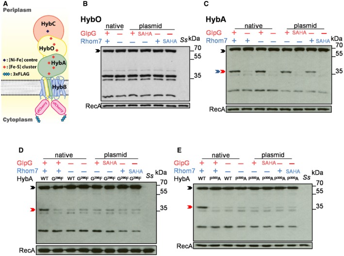
Schematic of HybA and HybO fusions.
Western blot analysis (probing with an anti‐FLAG mAb) to detect cleavage of HybO with (+)/without (−) chromosomal (native) or pBAD33‐encoded rhomboids (plasmid).
Western blot analysis to detect cleavage of HybA with (+)/without (−) chromosomal (native) or pBAD33‐encoded rhomboids (plasmid).
Western blot analysis to detect cleavage of HybAG296F by endogenous or pBAD33‐encoded rhomboid.
Western blot analysis to detect cleavage of HybAP300A by endogenous or pBAD33‐encoded rhomboid.
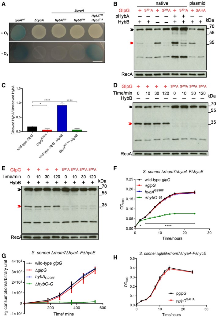
Bacterial two‐hybrid analysis with HybA and/or HybB fused chromosomally with the T25 and T18 domains of B. pertussis CyaA, respectively, in the absence of endogenous CyaA. Bacteria were grown on LB agar containing 20 μg/ml X‐gal at 37°C for 14 h in the presence/absence of O2. Scale bar, 1 cm.
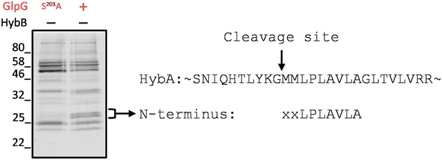
Western blot analysis to detect HybA cleavage in S. sonnei Δrhom7 by wild‐type (+) or inactive (S201A) GlpG expressed chromosomally (native) or from pUC19 (plasmid) with (+)/without (−) HybB. HybA was expressed from its native locus or a plasmid (pHybA).
Quantification of the ratio of cleaved/uncleaved HybA in strains with (+) or without (−) HybB. Mean ± S.D. of three experiments. *P < 0.05; ****P < 0.0001 (one‐way ANOVA).
Western blot analysis to detect GlpG‐mediated cleavage of native C‐terminally sfCherry‐3xFLAG‐tagged HybA at indicated times after blocking protein translation by the addition of chloramphenicol at T0 in the absence of HybB (−).
Western blot analysis to detect GlpG‐mediated cleavage of tagged HybA at times after blocking protein translation by the addition of chloramphenicol at T0 in the presence of HybB (+).
Growth of bacteria lacking hydrogenases (ΔhyaA‐F ΔhycE +/− ΔhybO‐G) or GlpG (ΔglpG), or expressing uncleavable HybA (hybA G296F) in 5% H2
H2 consumption by S. sonnei. Bacteria were grown aerobically in LB overnight and then diluted into M9 minimal media supplemented with 0.5% fumarate, 12.5 μg/ml nicotinic acid and 0.2% casamino acids in a sealed glass chromatography vial. The headspace was purged with 10% H2/90% argon, and cultures were incubated at 37 °C with shaking at 180 rpm for 9 h. H2 in the headspace was sampled and measured by gas chromatography.
Shigella sonnei growth in 10% H2 with plasmid‐expressed wild‐type or non‐functional (pglpG SAHA) GlpG.
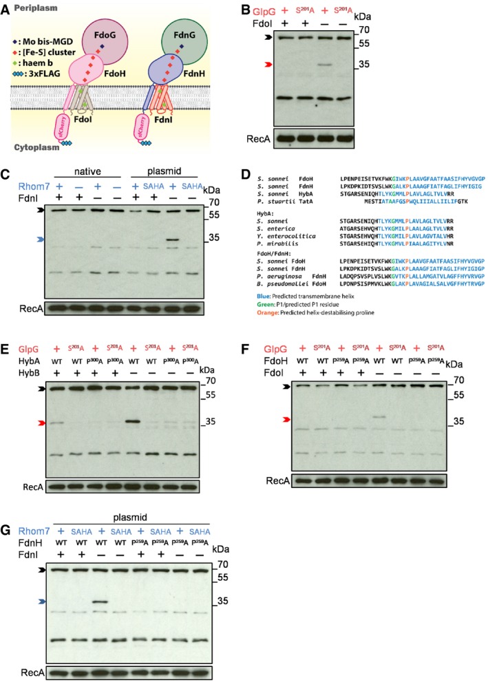
Schematic of FdoH and FdnH fusions.
Western blot analysis (probing with an anti‐FLAG mAb) to detect cleavage of FdoH cleavage in S. sonnei Δrhom7 with (+)/without (−) FdoI.
Western blot analysis to detect cleavage of FdnH in S. sonnei ΔglpG with (+) or without (−) chromosomal Rhom7 (native) or wild‐type (+)/inactive (SAHA) Rhom7 expressed from pBAD33 in S. sonnei ΔglpGΔrhom7 with (+) or without (−) FdnI.
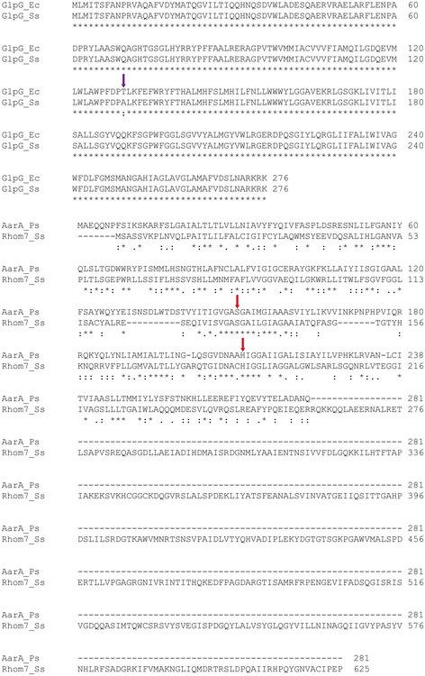
Alignments of HybA, FdoH and FdnH in S. sonnei with Providencia stuartii TatA and with their homologues from phylogenetically diverse organisms (S. sonnei, Salmonella enterica, Yersinia enterocolitica, Proteus mirabilis, Pseudomonas aeruginosa and Burkholderia pseudomallei) highlighting conserved glycine (green) and proline (orange) residues.
Western blot analysis to detect cleavage of wild‐type (WT) or uncleavable (P300A) HybA by active (+) or inactive (S201A) chromosomal GlpG in the presence (+) or absence (−) of HybB.
Western blot analysis to detect cleavage of wild‐type (WT) or modified (P259A) FdoH by active (+) or inactive (S201A) chromosomal GlpG in the presence (+) or absence (−) of FdoI.
Western blot analysis to detect cleavage of wild‐type (WT) or modified (P259A) FdnH with active (+) or inactive (SAHA) Rhom7 expressed from pBAD33 with (+) or without (−) FdnI.
- A
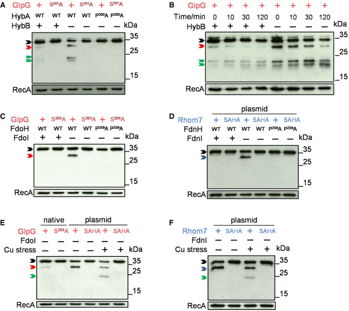
Western blot analysis (probing with an anti‐V5 mAb) to detect degradation of N‐terminally V5‐tagged wild‐type (WT) or modified (P300A) HybA in S. sonnei Δrhom7 chromosomally expressing wild‐type (+) or inactive (S201A) GlpG with (+) or without (−) HybB.
- B
Degradation of V5‐tagged HybA at times after blocking protein translation at T0 in the presence (+) or absence (−) of HybB.
- C
Western blot analysis of N‐terminally V5‐tagged wild‐type (WT) or modified (P259A) FdoH in S. sonnei Δrhom7 chromosomally expressing wild‐type (+) or inactive (S201A) GlpG with (+)/without (−) FdoI.
- D
Western blot analysis of N‐terminally V5‐tagged wild‐type (WT) or modified (P259A) FdnH in S. sonnei Δrhom7 with wild‐type (+) or inactive (SAHA) Rhom7 expressed from pBAD33 with (+)/without (−) FdnI.
- E, F
Degradation of N‐terminally V5‐tagged wild‐type (WT) or modified (P259A) FdoH (E) or FdnH (F) in S. sonnei Δrhom7 with wild‐type (+) or inactive (S201A, SAHA) GlpG expressed chromosomally (native) or from pUC19 (plasmid) without FdoI or FdnI (−), respectively, +/− exposure to 400 μM CuCl2 for 30 min.
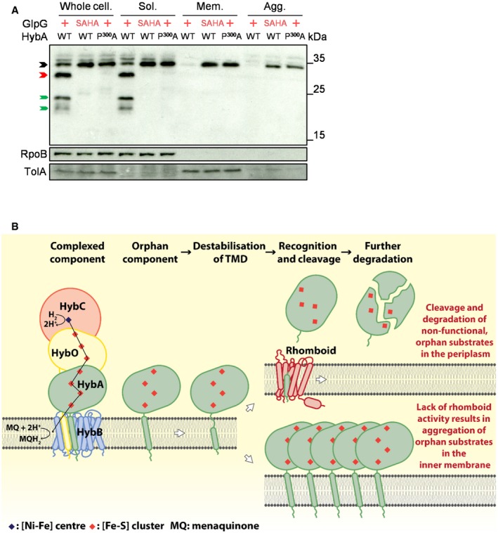
Western blot analysis probing the localisation and status of plasmid‐encoded N‐terminally V5‐tagged wild‐type (WT) or modified (P300A) HybA in S. sonnei Δrhom7ΔhybB with wild‐type (+) or inactive (SAHA) GlpG expressed from pUC19. Whole‐cell lysate (Whole cell.), Soluble (Sol.), Membrane (detergent‐solubilised, Mem.) and the Aggregate (Agg.) fractions are shown. HybA that is uncleaved, cleaved only by GlpG and further degraded post‐GlpG cleavage is marked by black, red and green arrows, respectively.
Model of rhomboid‐mediated quality control by selectively targeting orphan components of multiprotein respiratory complexes.
Comment in
-
Derlins with scissors: primordial ERAD in bacteria.EMBO J. 2020 May 18;39(10):e105012. doi: 10.15252/embj.2020105012. Epub 2020 Apr 27. EMBO J. 2020. PMID: 32338387 Free PMC article.
References
-
- Akiyama Y, Kihara A, Ito K (1996) Subunit a of proton ATPase F0 sector is a substrate of the FtsH protease in Escherichia coli . FEBS Lett 399: 26–28 - PubMed
Publication types
MeSH terms
Substances
Grants and funding
LinkOut - more resources
Full Text Sources
Other Literature Sources
