

Positron Emission Tomography Imaging With [18F]flortaucipir and Postmortem Assessment of Alzheimer Disease Neuropathologic Changes
- PMID: 32338734
- PMCID: PMC7186920
- DOI: 10.1001/jamaneurol.2020.0528
Positron Emission Tomography Imaging With [18F]flortaucipir and Postmortem Assessment of Alzheimer Disease Neuropathologic Changes
Erratum in
-
Error in Figure 3.JAMA Neurol. 2023 Aug 1;80(8):873. doi: 10.1001/jamaneurol.2023.1911. JAMA Neurol. 2023. PMID: 37306990 Free PMC article. No abstract available.
Abstract
Importance: Positron emission tomography (PET) may increase the diagnostic accuracy and confirm the underlying neuropathologic changes of Alzheimer disease (AD).
Objective: To determine the accuracy of antemortem [18F]flortaucipir PET images for predicting the presence of AD-type tau pathology at autopsy.
Design, setting, and participants: This diagnostic study (A16 primary cohort) was conducted from October 2015 to June 2018 at 28 study sites (27 in US sites and 1 in Australia). Individuals with a terminal illness who were older than 50 years and had a projected life expectancy of less than 6 months were enrolled. All participants underwent [18F]flortaucipir PET imaging, and scans were interpreted by 5 independent nuclear medicine physicians or radiologists. Supplemental autopsy [18F]flortaucipir images and pathological samples were also collected from 16 historically collected cases. A second study (FR01 validation study) was conducted from March 26 to April 26, 2019, in which 5 new readers assessed the original PET images for comparison to autopsy.
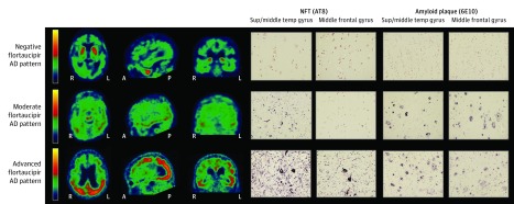
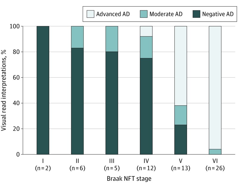
Main outcomes and measures: [18F]flortaucipir PET images were visually assessed and compared with immunohistochemical tau pathology. An AD tau pattern of flortaucipir retention was assessed for correspondence with a postmortem B3-level (Braak stage V or VI) pathological pattern of tau accumulation and to the presence of amyloid-β plaques sufficient to meet the criteria for high levels of AD neuropathological change. Success was defined as having at least 3 of the 5 readers above the lower bounds of the 95% CI for both sensitivity and specificity of 50% or greater.
Results: A total of 156 patients were enrolled in the A16 study and underwent [18F]flortaucipir PET imaging. Of these, 73 died during the study, and valid autopsies were performed for 67 of these patients. Three autopsies were evaluated as test cases and removed from the primary cohort (n = 64). Of the 64 primary cohort patients, 34 (53%) were women and 62 (97%) were white; mean (SD) age was 82.5 (9.6) years; and 49 (77%) had dementia, 1 (2%) had mild cognitive impairment, and 14 (22%) had normal cognition. Prespecified success criteria were met for the A16 primary cohort. The flortaucipir PET scans predicted a B3 level of tau pathology, with sensitivity ranging from 92.3% (95% CI, 79.7%-97.3%) to 100.0% (95% CI, 91.0%-100.0%) and specificity ranging from 52.0% (95% CI, 33.5%-70.0%) to 92.0% (95% CI, 75.0%-97.8%). A high level of AD neuropathological change was predicted with sensitivity of 94.7% (95% CI, 82.7%-98.5%) to 100.0% (95% CI, 90.8%-100.0%) and specificity of 50.0% (95% CI, 32.1%-67.9%) to 92.3% (95% CI, 75.9%-97.9%). The FR01 validation study also met prespecified success criteria. Addition of the supplemental autopsy data set and 3 test cases, which comprised a total of 82 patients and autopsies for both the A16 and FR01 studies, resulted in improved specificity and comparable overall accuracy. Among the 156 enrolled participants, 14 (9%) experienced at least 1 treatment-emergent adverse event.
Conclusions and relevance: This study's findings suggest that PET imaging with [18F]flortaucipir could be used to identify the density and distribution of AD-type tau pathology and the presence of high levels of AD neuropathological change, supporting a neuropathological diagnosis of AD.
Conflict of interest statement
Figures

Comment in
-
Imaging Tau Pathology-The Next Step.JAMA Neurol. 2020 Jul 1;77(7):796-797. doi: 10.1001/jamaneurol.2020.0520. JAMA Neurol. 2020. PMID: 32338712 No abstract available.
References
-
- Masters CL, Multhaup G, Simms G, Pottgiesser J, Martins RN, Beyreuther K. Neuronal origin of a cerebral amyloid: neurofibrillary tangles of Alzheimer’s disease contain the same protein as the amyloid of plaque cores and blood vessels. EMBO J. 1985;4(11):2757-2763. doi: 10.1002/j.1460-2075.1985.tb04000.x - DOI - PMC - PubMed
Publication types
MeSH terms
Substances
Grants and funding
- P50 AG016574/AG/NIA NIH HHS/United States
- R01 AG041851/AG/NIA NIH HHS/United States
- R01 AG034676/AG/NIA NIH HHS/United States
- R01 NS089757/NS/NINDS NIH HHS/United States
- P01 AG066597/AG/NIA NIH HHS/United States
- P01 AG019724/AG/NIA NIH HHS/United States
- P30 AG019610/AG/NIA NIH HHS/United States
- R21 NS094489/NS/NINDS NIH HHS/United States
- P30 AG062422/AG/NIA NIH HHS/United States
- R01 AG011378/AG/NIA NIH HHS/United States
- R01 AG054449/AG/NIA NIH HHS/United States
- P50 AG023501/AG/NIA NIH HHS/United States
- R01 NS089544/NS/NINDS NIH HHS/United States
- U01 AG006786/AG/NIA NIH HHS/United States
- R01 NS097495/NS/NINDS NIH HHS/United States
LinkOut - more resources
Full Text Sources
Other Literature Sources
Medical
Miscellaneous
