. 2020 Sep;296(3):E156-E165.
doi: 10.1148/radiol.2020201491. Epub 2020 Apr 27.

Artificial Intelligence Augmentation of Radiologist Performance in Distinguishing COVID-19 from Pneumonia of Other Origin at Chest CT

Affiliations

Artificial Intelligence Augmentation of Radiologist Performance in Distinguishing COVID-19 from Pneumonia of Other Origin at Chest CT

Harrison X Bai et al. Radiology. 2020 Sep.

Erratum in

Abstract

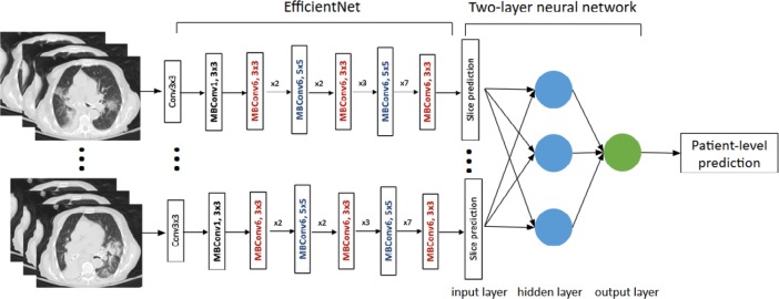
Background Coronavirus disease 2019 (COVID-19) and pneumonia of other diseases share similar CT characteristics, which contributes to the challenges in differentiating them with high accuracy. Purpose To establish and evaluate an artificial intelligence (AI) system for differentiating COVID-19 and other pneumonia at chest CT and assessing radiologist performance without and with AI assistance. Materials and Methods A total of 521 patients with positive reverse transcription polymerase chain reaction results for COVID-19 and abnormal chest CT findings were retrospectively identified from 10 hospitals from January 2020 to April 2020. A total of 665 patients with non-COVID-19 pneumonia and definite evidence of pneumonia at chest CT were retrospectively selected from three hospitals between 2017 and 2019. To classify COVID-19 versus other pneumonia for each patient, abnormal CT slices were input into the EfficientNet B4 deep neural network architecture after lung segmentation, followed by a two-layer fully connected neural network to pool slices together. The final cohort of 1186 patients (132 583 CT slices) was divided into training, validation, and test sets in a 7:2:1 and equal ratio. Independent testing was performed by evaluating model performance in separate hospitals. Studies were blindly reviewed by six radiologists without and then with AI assistance. Results The final model achieved a test accuracy of 96% (95% confidence interval [CI]: 90%, 98%), a sensitivity of 95% (95% CI: 83%, 100%), and a specificity of 96% (95% CI: 88%, 99%) with area under the receiver operating characteristic curve of 0.95 and area under the precision-recall curve of 0.90. On independent testing, this model achieved an accuracy of 87% (95% CI: 82%, 90%), a sensitivity of 89% (95% CI: 81%, 94%), and a specificity of 86% (95% CI: 80%, 90%) with area under the receiver operating characteristic curve of 0.90 and area under the precision-recall curve of 0.87. Assisted by the probabilities of the model, the radiologists achieved a higher average test accuracy (90% vs 85%, Δ = 5, P < .001), sensitivity (88% vs 79%, Δ = 9, P < .001), and specificity (91% vs 88%, Δ = 3, P = .001). Conclusion Artificial intelligence assistance improved radiologists' performance in distinguishing coronavirus disease 2019 pneumonia from non-coronavirus disease 2019 pneumonia at chest CT. © RSNA, 2020 Online supplemental material is available for this article.


Figures

Diagram illustrating patient inclusion and exclusion. Abbreviations:
RIH, Rhode Island Hospital; HUP, Hospital of the University of Pennsylvania;
AI, artificial intelligence; RT-PCR, reverse transcriptase polymerase chain
reaction.
Figure 1.
Diagram illustrating patient inclusion and exclusion. Abbreviations: RIH, Rhode Island Hospital; HUP, Hospital of the University of Pennsylvania; AI, artificial intelligence; RT-PCR, reverse transcriptase polymerase chain reaction.
Flow diagram illustrating our AI model for distinguishing COVID-19
from non-COVID-19 pneumonia. Abbreviations: ROC AUC: Receiver Operator
Characteristics Area Under the Curve; PR AUC: Precision Recall area under
curve.
Figure 2.
Flow diagram illustrating our AI model for distinguishing COVID-19 from non-COVID-19 pneumonia. Abbreviations: ROC AUC: Receiver Operator Characteristics Area Under the Curve; PR AUC: Precision Recall area under curve.
COVID-19 Classification Neural Network Model.
Figure 3:
COVID-19 Classification Neural Network Model.
ROC curve of deep neural network on the test set compared to
radiologist performance. ROC = receiver operating curve.
Figure 4.
ROC curve of deep neural network on the test set compared to radiologist performance. ROC = receiver operating curve.
Representative slices corresponding to Grad-CAM images on the test
set.
Figure 5.
Representative slices corresponding to Grad-CAM images on the test set.
Representative cases that the majority of radiologists misclassified.
A-C (top row, left to right): COVID-19 pneumonia. Our model correctly
classified all three cases. A. 4/6 radiologists (radiologists 3-6) said it
was non-COVID-19. With AI assistance, 2/6 radiologists (radiologists 5 and
6) continued to say it was non-COVID-19. B. 4/6 radiologists (radiologists
3-6) said it was non-COVID-19. With AI assistance, 3/6 radiologists
(radiologists 3-5) continued to say it was non-COVID-19. C. 4/6 radiologists
(radiologists 2 and 4-6) said it was non-COVID-19. With AI assistance, 1/6
radiologist (radiologist 2) continued to say it was non-COVID-19. D-F
(bottom row, left to right): Non-COVID-19 pneumonia. Our model correctly
classified D and E. D. 5/6 radiologists (radiologists 1-5) said it was
COVID-19. With AI assistance, all 5/6 radiologists continued to say it was
COVID-19. E. 4/6 radiologists (radiologists 1, 2, 4, and 6) said it was
COVID-19. With AI assistance, 3/6 radiologists (radiologists 1, 2, and 4)
continued to say it was COVID-19. F. 4/6 radiologists (radiologists 1-3 and
6) said it was COVID-19. With AI assistance, 5/6 radiologists (radiologists
1-4 and 6) said it was COVID-19.
Figure 6.
Representative cases that the majority of radiologists misclassified. A-C (top row, left to right): COVID-19 pneumonia. Our model correctly classified all three cases. A. 4/6 radiologists (radiologists 3-6) said it was non-COVID-19. With AI assistance, 2/6 radiologists (radiologists 5 and 6) continued to say it was non-COVID-19. B. 4/6 radiologists (radiologists 3-6) said it was non-COVID-19. With AI assistance, 3/6 radiologists (radiologists 3-5) continued to say it was non-COVID-19. C. 4/6 radiologists (radiologists 2 and 4-6) said it was non-COVID-19. With AI assistance, 1/6 radiologist (radiologist 2) continued to say it was non-COVID-19. D-F (bottom row, left to right): Non-COVID-19 pneumonia. Our model correctly classified D and E. D. 5/6 radiologists (radiologists 1-5) said it was COVID-19. With AI assistance, all 5/6 radiologists continued to say it was COVID-19. E. 4/6 radiologists (radiologists 1, 2, 4, and 6) said it was COVID-19. With AI assistance, 3/6 radiologists (radiologists 1, 2, and 4) continued to say it was COVID-19. F. 4/6 radiologists (radiologists 1-3 and 6) said it was COVID-19. With AI assistance, 5/6 radiologists (radiologists 1-4 and 6) said it was COVID-19.

Comment in

Similar articles

Cited by

References

    1. Bai Y, Yao L, Wei T, Tian F, Jin DY, Chen L, et al. . Presumed Asymptomatic Carrier Transmission of COVID-19 . JAMA . 2020. . Epub 2020/02/23. doi: 10.1001/jama.2020.2565. PubMed PMID: 32083643; PubMed Central PMCID: PMCPMC7042844 . - PMC - PubMed
    1. Qiu H, Wu J, Hong L, Luo Y, Song Q, Chen D. . Clinical and epidemiological features of 36 children with coronavirus disease 2019 (COVID-19) in Zhejiang, China: an observational cohort study . The Lancet Infectious Diseases . 2020. . - PMC - PubMed
    1. Huang C, Wang Y, Li X, Ren L, Zhao J, Hu Y, et al. . Clinical features of patients infected with 2019 novel coronavirus in Wuhan, China . Lancet . 2020. ; 395 ( 10223 ): 497 - 506 . Epub 2020/01/28. doi: 10.1016/S0140-6736(20)30183-5. PubMed PMID: 31986264 . - PMC - PubMed
    1. Wang D, Hu B, Hu C, Zhu F, Liu X, Zhang J, et al. . Clinical characteristics of 138 hospitalized patients with 2019 novel coronavirus–infected pneumonia in Wuhan, China . Jama . 2020. . - PMC - PubMed
    1. Bai HX, Hsieh B, Xiong Z, Halsey K, Choi JW, Tran TML, et al. . Performance of radiologists in differentiating COVID-19 from viral pneumonia on chest CT . Radiology . 2020. : 200823 -. - PMC - PubMed

MeSH terms