. 2020 Apr 27;16(4):e1008721.
doi: 10.1371/journal.pgen.1008721. eCollection 2020 Apr.

Mutations in SPATA13/ASEF2 cause primary angle closure glaucoma

Affiliations

Mutations in SPATA13/ASEF2 cause primary angle closure glaucoma

Naushin H Waseem et al. PLoS Genet. .

Abstract

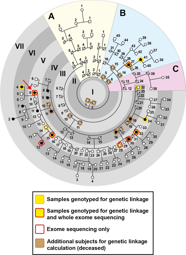
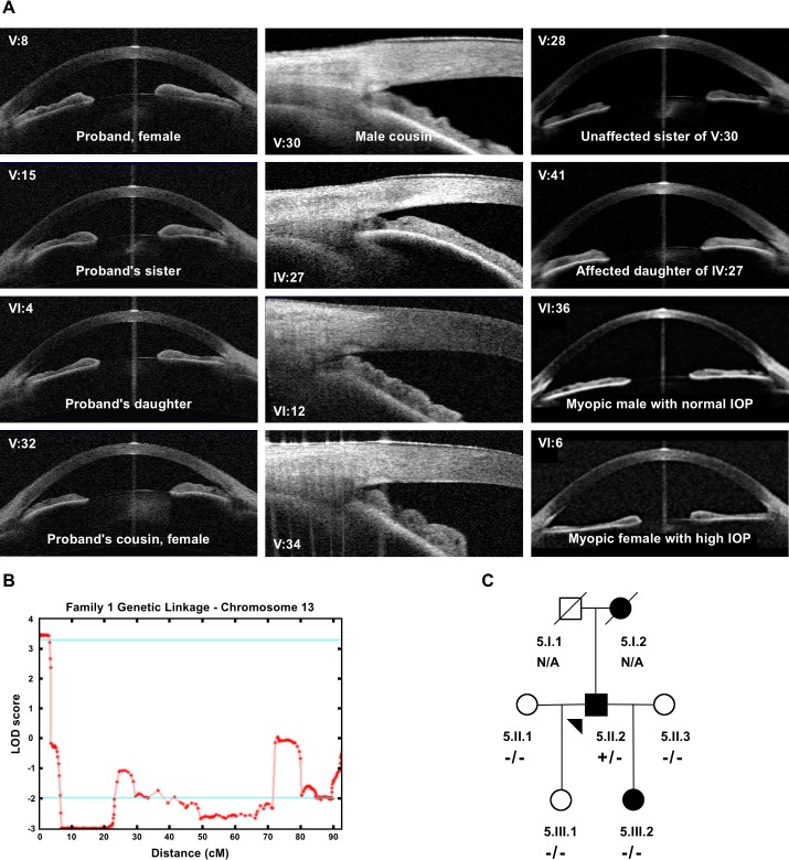
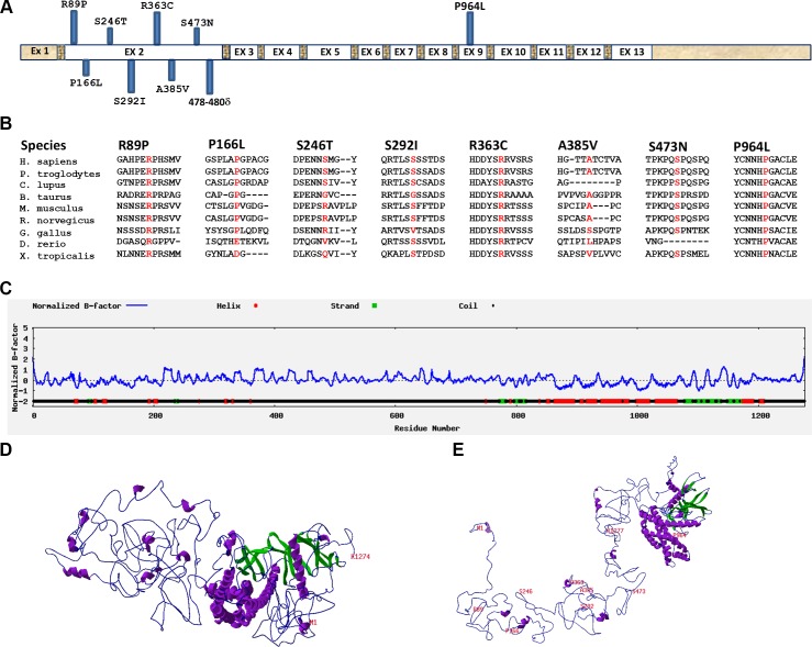
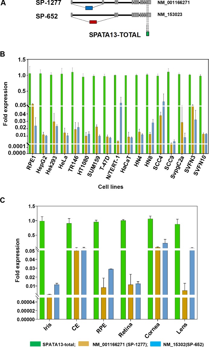
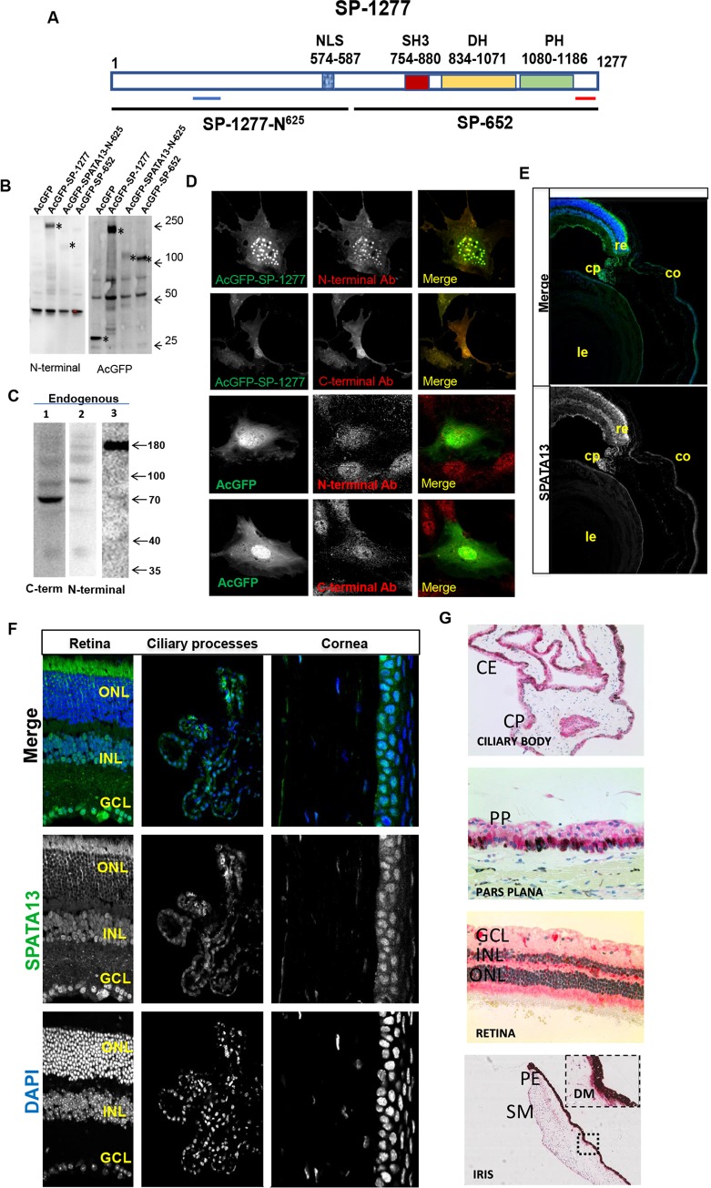
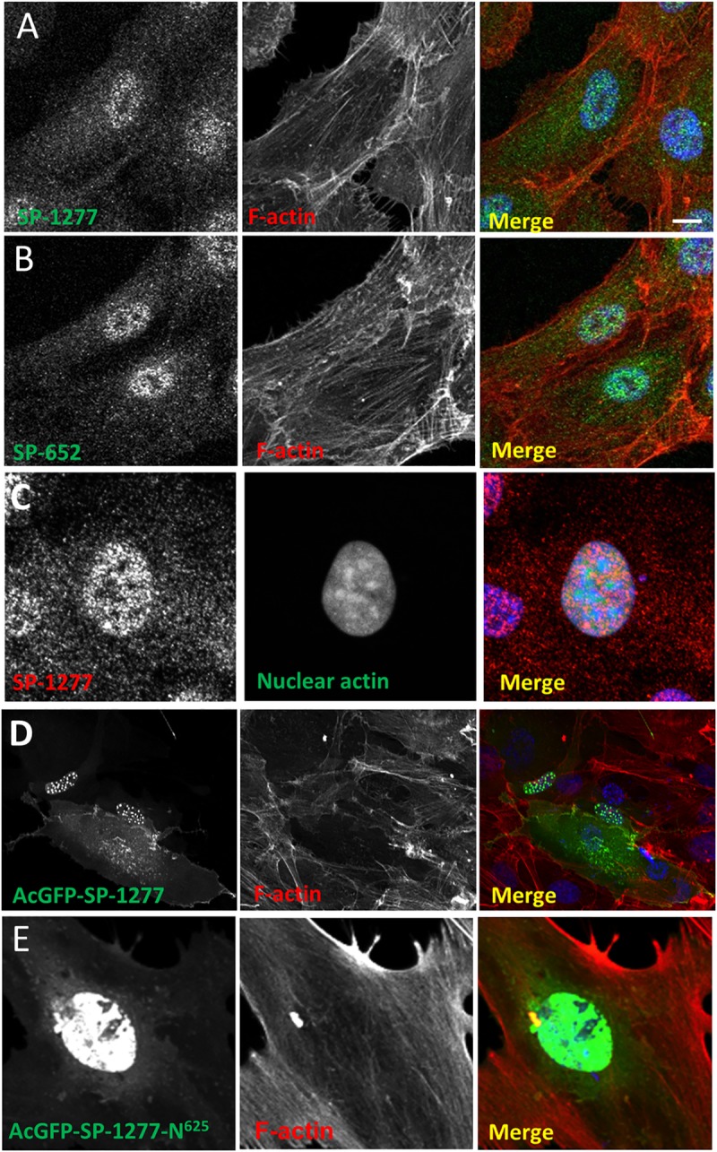
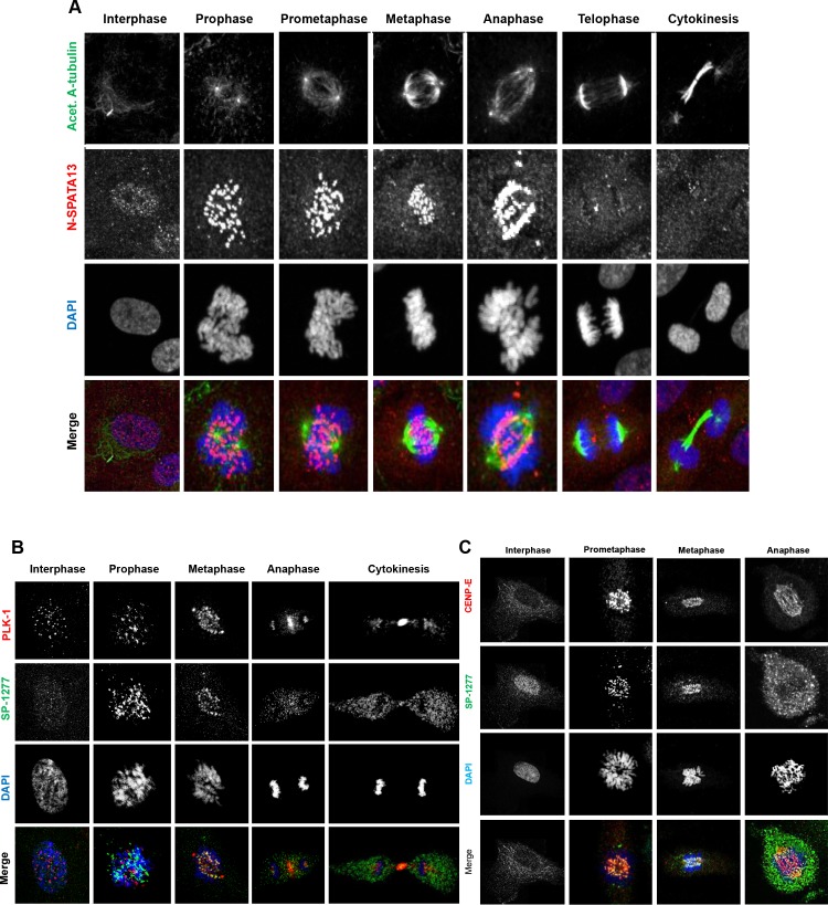
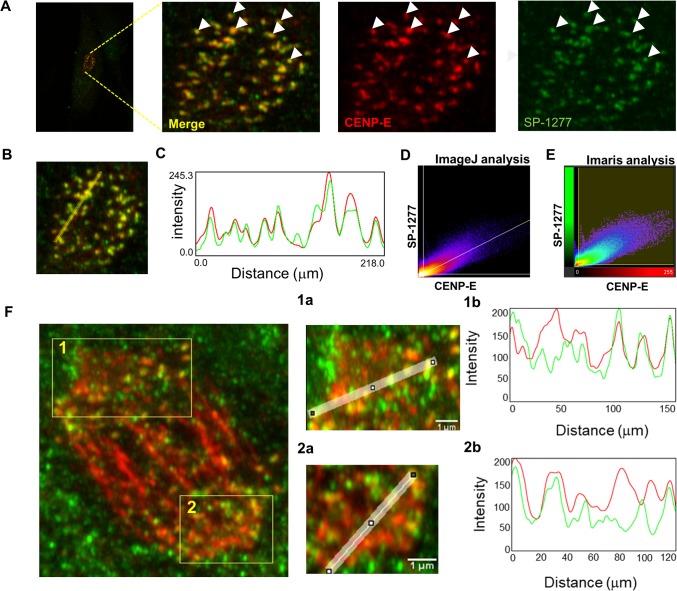
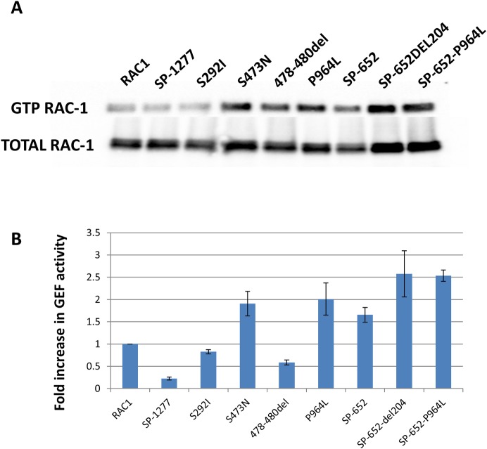
Current estimates suggest 50% of glaucoma blindness worldwide is caused by primary angle-closure glaucoma (PACG) but the causative gene is not known. We used genetic linkage and whole genome sequencing to identify Spermatogenesis Associated Protein 13, SPATA13 (NM_001166271; NP_001159743, SPATA13 isoform I), also known as ASEF2 (Adenomatous polyposis coli-stimulated guanine nucleotide exchange factor 2), as the causal gene for PACG in a large seven-generation white British family showing variable expression and incomplete penetrance. The 9 bp deletion, c.1432_1440del; p.478_480del was present in all affected individuals with angle-closure disease. We show ubiquitous expression of this transcript in cell lines derived from human tissues and in iris, retina, retinal pigment and ciliary epithelia, cornea and lens. We also identified eight additional mutations in SPATA13 in a cohort of 189 unrelated PACS/PAC/PACG samples. This gene encodes a 1277 residue protein which localises to the nucleus with partial co-localisation with nuclear speckles. In cells undergoing mitosis SPATA13 isoform I becomes part of the kinetochore complex co-localising with two kinetochore markers, polo like kinase 1 (PLK-1) and centrosome-associated protein E (CENP-E). The 9 bp deletion reported in this study increases the RAC1-dependent guanine nucleotide exchange factors (GEF) activity. The increase in GEF activity was also observed in three other variants identified in this study. Taken together, our data suggest that SPATA13 is involved in the regulation of mitosis and the mutations dysregulate GEF activity affecting homeostasis in tissues where it is highly expressed, influencing PACG pathogenesis.


Conflict of interest statement

The authors have declared that no competing interests exist.

Figures

Fig 1
Fig 1. Pedigree of Family 1.
The proband is V:8 (red arrow) and this pedigree shows the relationship of the 62 individuals examined (see S1 Table for detailed phenotype). There was no male-to-male transmission observed, and the only affected male was V:30, a first cousin of the proband. From branch B, two out of nine individuals examined were affected. There were no individuals examined from branches A and C affected with angle-closure disease. Twelve subjects, four of whom were affected first cousins, were genotyped for genetic linkage analysis, and some were analysed in greater detail by exome and whole genome sequencing. It is suspected that the phenotype in VII:2 and VI:7 is caused by another genetic defect as it is very likely that their disease is inherited from the married in spouse (V:11).
Fig 2
Fig 2. Clinical features and linkage analysis of Family 1.
(A) Plateau iris configuration was observed for all nine clearly affected individuals V:8, V:15, VI:4, V:32, V:30, IV:27, VI:12 and V:34, V:41) on gonioscopy. In the first two columns, closed anterior chamber drainage angles are observed. The middle column shows high resolution AS-OCT angle imaging, where the bulky ciliary body typical of plateau iris is observed. Individual IV:27 is the oldest and most severely affected PACG individual, showing some additional iris convexity. Individuals V:28, VI:36 and VI:6 have open drainage angles and are shown for comparison. (B) Maximum multipoint LOD score of 3.5 achieved at chromosome 13 by Superlink-Online SNP-1.1. (C) Pedigree of the family 5 identified with the 9 bp deletion. The proband, 5.II.2, shown by an arrowhead carry the deletion. His mother, 5.I.2 was affected but no DNA was available for analysis. Proband has an affected daughter, 5.III.2, but her eye phenotype is different to his. Her mother, 5:II.3, has narrow but open angles and was hyperopic and it is therefore suspected that the daughter has inherited her disease from the mother.
Fig 3
Fig 3. Location and predicted effect of mutations in SPATA13 gene on protein conformation.
(A) Structure of SPATA13 gene structure and location of 8 mutations in exon 2 and the 9th mutation in exon 9. (B) Conservation of amino acids mutated in PACG in 9 different species. (C) Secondary structure prediction for the amino acid sequence of SPATA13 polypeptide. (D) Predicted tertiary structure of SP-1277 containing the 9bp (p. 478-480del) deletion. (E) Wildtype SP-1277 showing the location of all the missense variants.
Fig 4
Fig 4. Expression of different SPATA13 transcripts in human cell lines and eye tissues.
(A) Schematic representation of the transcripts of different isoforms of SPATA13. The two isoform SP-1277 and SP-652 isoforms are generated by alternative splicing of exons toward the N-terminal. The position of qPCR primers used to differentiate the 2 transcripts are also shown. (B) Expression of SPATA13 transcripts by qPCR in 17 different cell lines derived from human eye, breast, liver, skin, head and neck, cervix and kidney. (C) Expression of SP-1277 and SP-652 transcripts in human iris, ciliary epithelium, retinal pigment epithelium, retina, cornea and lens. The green bars represent SPATA13-Total whereas the blue bars represent SP-1277 and the red bars represent SP-652.
Fig 5
Fig 5. Characterisation of SP-1277 isoform.
(A) Schematic representation of the domain structure of SP-1277 isoform. Light blue bar shows the epitope for the N-terminal antibodies and the red bar shows epitope for the C-terminal antibodies. The nuclear localisation signal in the terminal region SP-1277N625is shown by a hashed blue bar. The boundary of SP-1277-N625 and SP-652 are shown two discontinuous lines. The SH3, DBL homology (DH) and PH domains are shown in SP-652. (B) Western blot analysis of the SPATA13 and its fragments analysed by N-terminus (ab122627) and anti-AcGFP antibodies. AcGFP fusion protein of SP-1277, SP-1277-N625 and the SP-652 were expressed in HT1080 cells, total cell lysate was separated on 4–15% SDS polyacrylamide gel, transferred on nitrocellulose membrane and probed with different antibodies. Specific reactivity with protein bands is shown by asterisks. (C) Western blot of total RPE-1 lysate probed with the C-terminus (lane 1) and N-terminal (lane 2, ThermoFisher; lane 3, Abcam) antibodies. (D) Reactivity of N- and C-terminal antibodies with RPE-1 cells overexpressing SP-1277 compared with AcGFP control. Immuohistochemical reactivity of murine eye tissues (E, F) and human eye tissues (G) with the N-terminus antibody from Abcam (ab122627). CE = ciliary epithelium, CP = ciliary process, PP = pars plana, GCL = ganglion cell layer, INL = inner nuclear layer, ONL = outer nuclear layer, SM = sphincter muscle, DM = dilator muscle, PE = posterior muscle. Re = retina, Co = cornea, Le = lens.
Fig 6
Fig 6. Endogenous and ectopic expression of SPATA13 isoforms.
Human RPE-1 cells were immunostained with SP-1277 (N-terminus antibody, A) and SP-652 (C-terminus antibody, B) and co-stained for endogenous F-actin (Alexa Fluor 594 conjugated phalloidin). (C) To detect nuclear actin, RPE-1 cells were transfected with nuclear actin chromobody (nAC) probe construct [55] and were counterstained with N-terminal specific SP-1277 antibody. RPE-1 cells were transiently transfected with AcGFP-tagged SP-1277 (D), and SP-1277-N625(E) respectively, both counterstained for F-actin. AcGFP-SP-1277 localised to nuclear speckles with a diffuse but variable cytoplasmic signal (S), AcGFP-SPATA13-N625 (E) only showed nuclear staining. Nuclei were stained with DAPI (blue). Scale bar = 10 μM.
Fig 7
Fig 7. Endogenous SP-1277 isoform during mitosis.
Endogenous SP-1277 protein was localised in dividing RPE-1 cells, during mitosis using the N-terminus antibody and counterstained using anti-acetylated ⍺-tubulin antibody (A), or counter stained with two mitotic markers PLK-1 (B) and CENP-E (C). (A) SP-1277 was detected in the nucleus as speckles at G0, which intensified at the onset of prophase, aligning along the kinetochore at metaphase, and anaphase, before returning to its G0 localisation pattern. (B) SP-1277 globules show co-localisation with the kinetochore marker PLK-1 at prophase, metaphase and anaphase. At cytokinesis the PLK-1 staining is primarily localized at midbody between the dividing cell which also co-stains for SP-1277. (C) SP-1277 show strong co-localisation with another kinetochore marker CENP-E at prophase, prometaphase and anaphase. At anaphase the colocalization primarily is around the separated chromosomes. Nuclei were stained using DAPI (blue).
Fig 8
Fig 8. Colocalization analysis of SP-1277 with CENP-E at prometaphase and anaphase.
(A) At prometaphase SP-1277 (green) co-localises with CENPE-E (red) globules. To evaluate whether colocalization occurs, a line was drawn connecting different globules (B). The green and red intensity was measured using ImageJ in globules along the line and plotted (C). The data was analysed using Image J and Imaris softwares and showed colocalization of 84% (tM0.84) of CENP-E with SP-1277 and 75% (tM = 0.75) of SP-1277 with CENP-E. Fluorescence intensity data of two images were distributed linearly as shown in the scatterplots (Fig 8D & 8E). The colocalization of SP-1277 with CENP-E in anaphase is shown in (F). The regions of interest showing highest colocalization are labelled as 1 and 2. The degree of colocalization of the two molecules was analysed using ImageJ (1a, and 1b for region 1) and (2a and 2b for region 2) and gave a colocalization of more than 80%.
Fig 9
Fig 9. SPATA13 mutations induced GEF activity in SP-1277.
(A) RPE-1 cells were co-transfected with Flag-RAC-1 along with wildtype SP-1277, wildtype SP-652 or mutants (S292I, S473N, 478-480d, P964L) cloned in pLPCpuro_NAcGFP and after lysis the GTP-RAC-1 was immunoprecipitated with PAK-PBD beads and analysed on western blot using anti-Flag antibody. (B) Quantification of GEF activity of SP-1277 (wildtype and mutants). One way ANOVA was used to test the null hypothesis of no difference between GEF activity in mutants and wildtype, with there being a significant difference (p = 0.002).

References

    1. Quigley HA. Angle-closure glaucoma-simpler answers to complex mechanisms: LXVI Edward Jackson Memorial Lecture. Am J Ophthalmol. 2009;148(5):657–69 e1. Epub 2009/11/03. 10.1016/j.ajo.2009.08.009 . - DOI - PubMed
    1. Foster PJ, Buhrmann R, Quigley HA, Johnson GJ. The definition and classification of glaucoma in prevalence surveys. Br J Ophthalmol. 2002;86(2):238–42. Epub 2002/01/30. 10.1136/bjo.86.2.238 ; PMCID: PMC1771026. - DOI - PMC - PubMed
    1. Weinreb RN, Khaw PT. Primary open-angle glaucoma. The Lancet. 2004;363(9422):1711–20. 10.1016/s0140-6736(04)16257-0 - DOI - PubMed
    1. Quigley HA, Broman AT. The number of people with glaucoma worldwide in 2010 and 2020. Br J Ophthalmol. 2006;90(3):262–7. Epub 2006/02/21. 10.1136/bjo.2005.081224 ; PMCID: PMC1856963. - DOI - PMC - PubMed
    1. Alsbirk PH. Anterior chamber depth, genes and environment. A population study among long-term Greenland Eskimo immigrants in Copenhagen. Acta Ophthalmol (Copenh). 1982;60(2):223–4. Epub 1982/04/01. 10.1111/j.1755-3768.1982.tb08376.x . - DOI - PubMed

Publication types

MeSH terms

Substances

LinkOut - more resources