. 2020 Jun 12;432(13):3869-3880.
doi: 10.1016/j.jmb.2020.04.020. Epub 2020 Apr 25.

Targeted Killing of Pseudomonas aeruginosa by Pyocin G Occurs via the Hemin Transporter Hur

Affiliations

Targeted Killing of Pseudomonas aeruginosa by Pyocin G Occurs via the Hemin Transporter Hur

Iva Atanaskovic et al. J Mol Biol. .

Abstract

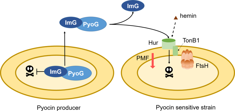
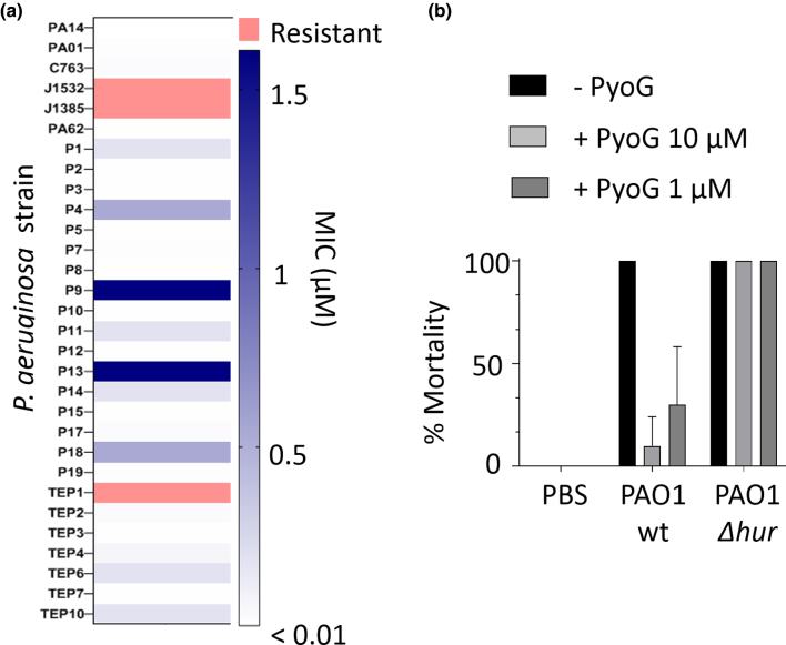
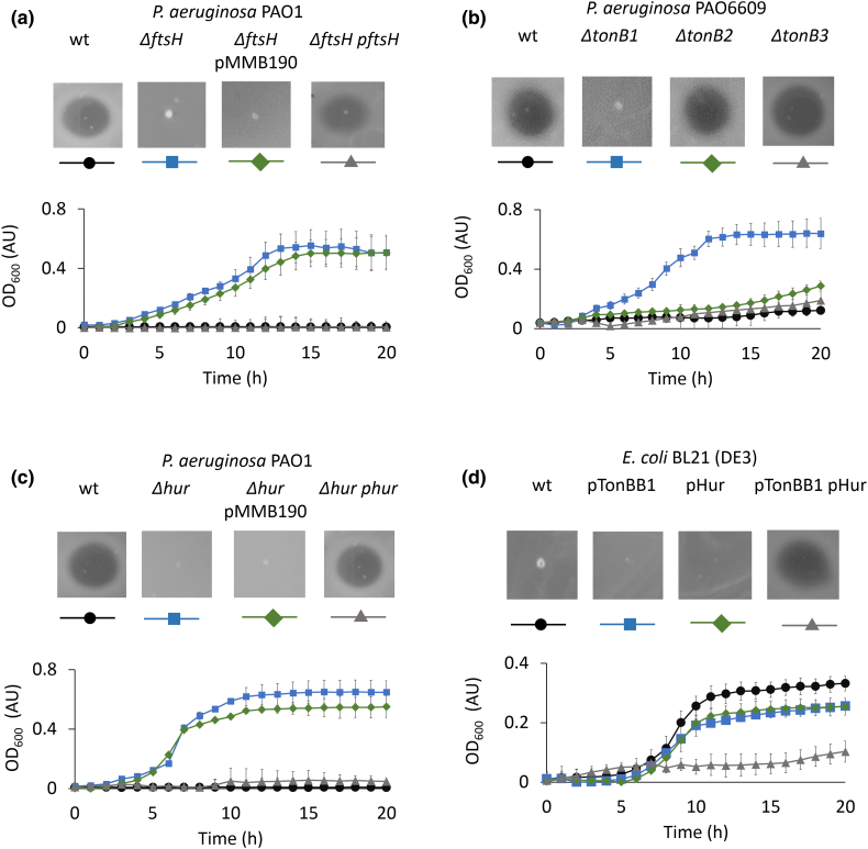
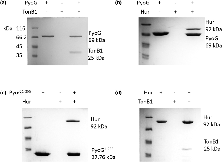
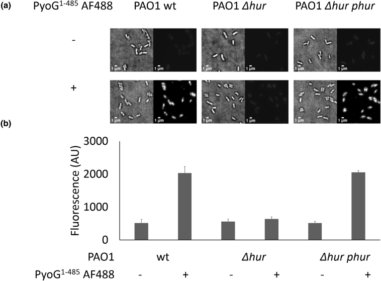
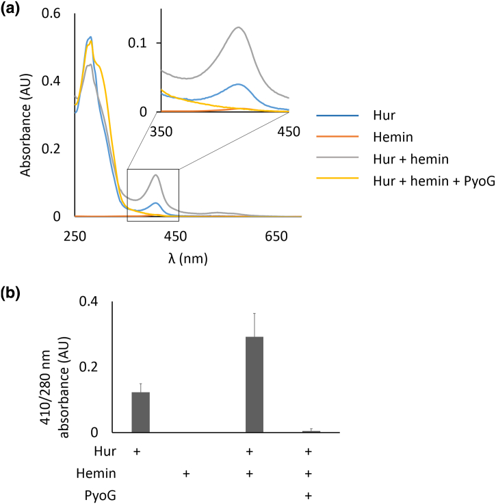
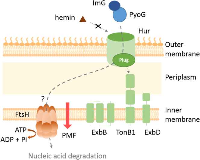
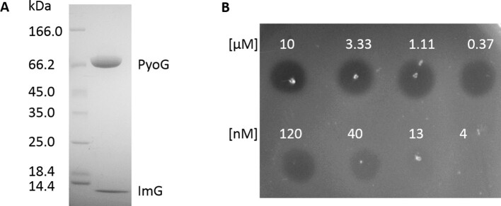
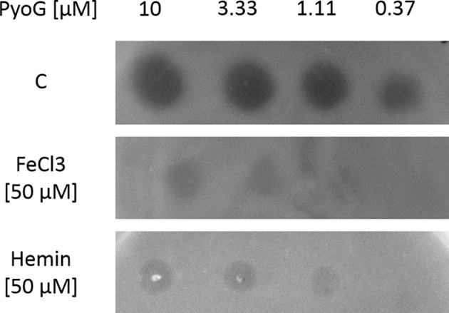
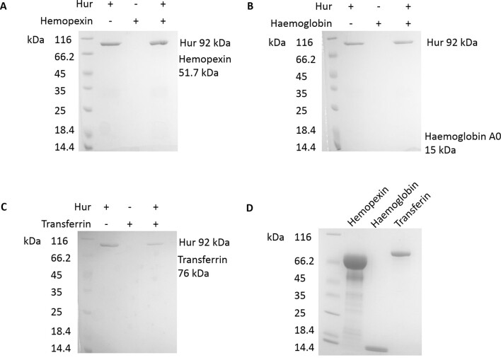
Pseudomonas aeruginosa is a priority pathogen for the development of new antibiotics, particularly because multi-drug-resistant strains of this bacterium cause serious nosocomial infections and are the leading cause of death in cystic fibrosis patients. Pyocins, bacteriocins of P. aeruginosa, are potent and diverse protein antibiotics that are deployed during bacterial competition. Pyocins are produced by more than 90% of P. aeruginosa strains and may have utility as last resort antibiotics against this bacterium. In this study, we explore the antimicrobial activity of a newly discovered pyocin called pyocin G (PyoG). We demonstrate that PyoG has broad killing activity against a collection of clinical P. aeruginosa isolates and is active in a Galleria mellonella infection model. We go on to identify cell envelope proteins that are necessary for the import of PyoG and its killing activity. PyoG recognizes bacterial cells by binding to Hur, an outer-membrane TonB-dependent transporter. Both pyocin and Hur interact with TonB1, which in complex with ExbB-ExbD links the proton motive force generated across the inner membrane with energy-dependent pyocin translocation across the outer membrane. Inner-membrane translocation of PyoG is dependent on the conserved inner-membrane AAA+ ATPase/protease, FtsH. We also report a functional exploration of the PyoG receptor. We demonstrate that Hur can bind to hemin in vitro and that this interaction is blocked by PyoG, confirming the role of Hur in hemin acquisition.

Keywords: FtsH; TonB-dependent transporter; bacteriocin; protein antibiotic; protein import.


Figures

Unlabelled Image
Graphical abstract
Figure 1
Figure 1
PyoG is active against clinical isolates of P. aeruginosa and can rescue G. mallonella from a lethal dose of P. aeruginosa PAO1. (a) PyoG MICs for a collection of P. aeruginosa clinical isolates. Resistant strains are shown in red, and MICs values are represented as shades of blue. Most strains are sensitive to the pyocin. (b) In vivo activity of PyoG against a lethal dose of P. aeruginosa PAO1 in the G. mallonella infection model. PAO1 Δhur is a PyoG-resistant mutant which lacks the receptor for the pyocin.
Figure 2
Figure 2
Plate and liquid killing assays with 10 μM PyoG reveal which cell envelope proteins are involved in its import. For OD600, mean of three biological replicates with standard deviations is shown. (a) FtsH, an inner-membrane AAA + ATPase/protease, is necessary for PyoG killing activity. P. aeruginosa PAO1 ΔftsH is resistant to PyoG and is unaffected by the introduction of the empty shuttle vector pMMB190 (ΔftsH pMMB190). However, transformation of PAO1 ΔftsH with pftsH complemented the ftsH deletion and restored sensitivity to PyoG. (b) TonB1, a protein that links the PMF generated on the inner membrane with translocation across the outer membrane, is necessary for PyoG killing activity. ΔtonB1 mutant of P. aeruginosa is resistant to PyoG, while ΔtonB2 and ΔtonB3 mutants are sensitive. (c) The killing activity of PyoG depends on an outer-membrane transporter, Hur. Deletion of hur induces resistance to the pyocin. Sensitivity can be restored if hur is complemented from a plasmid. Phur is hur cloned from PAO1 into pMMB190. (d) E. coli BL21 (DE3) is not sensitive to PyoG. PyoG sensitivity in this organism can be induced if transformed with both pTonBB1 and pHur. pTonBB1 is E. coli TonB1–102 translationally fused to P. aeruginosa TonB1201–342 and cloned into pACYCDuet-1 [6]. pHur is hur with the E. coli OmpF signal sequence, codon optimized for expression in E. coli and cloned into pET21d.
Figure 3
Figure 3
Pull-downs of PyoG translocon components. Proteins were mixed at equimolar concentrations and bound to nickel beads, which were then washed of unbound protein. Eluate content was analyzed on 12% SDS-PAGE gels. A protein marker is shown in the first lane on each gel. Eluate of the bait protein is in the second lane. Eluate of the prey protein is shown in the third lane and it verifies that the prey does not bind to beads on its own. Eluate for the mix of bait and prey is shown in the fourth lane. Positions of proteins with their molecular masses are labeled on the right side of the gel. (a) PyoG and the periplasmic region of TonB1 interact in vitro. (b) PyoG binds to Hur, confirming its involvement in the import of the pyocin. (c) The first 255 residues of PyoG, which are conserved among S1-group pyocins, bind to Hur. (d) Hur binds to the periplasmic region of TonB1, confirming that this is a TonB1-dependent transporter.
Figure 4
Figure 4
Hur is required for PyoG to target P. aeruginosa cells. (a) Fluorescent labeling of PAO1 wt, Δhur and Δhur complemented with Hur expressed from a plasmid (Δhur phur). PyoG1–483, conjugated to AF488 via a C-terminal cysteine, was used for labeling. This construct labels the wt strain, but there is loss of labeling if hur is deleted. Labeling is restored if hur is complemented from a plasmid. Representative micrographs for each strain are shown. (b) Average fluorescence intensities for 100 cells in the presence and absence of fluorescent PyoG. Mean of three biological replicates with standard deviations is shown.
Figure 5
Figure 5
Hur binds hemin, a source of iron for P. aeruginosa in mammalian hosts. Hemin binding to Hur can be blocked by PyoG. (a) Pull-down of hemin with 10 μM His-tagged Hur, in the presence and absence of 10 μM PyoG lacking a purification tag. Proteins were mixed with a 100 × molar excess of hemin and bound to nickel beads. Beads were then washed of unbound protein and hemin. Absorbance spectra of eluate were measured to detect changes in the 410-nm hemin peak (enlarged in the upper corner). Representative absorbance spectra are shown. The 410-nm peak is increased if Hur is exposed to excess hemin, and no hemin peak can be observed if Hur was mixed with PyoG. No considerable 410-nm peak in the protein free control, containing hemin only, confirms that unbound hemin was washed off the beads and makes no contribution to the 410-nm absorbance in the eluate. (b) Ratio between the hemin 410-nm peak and the protein 280-nm peak in the eluate indicates that Hur binds hemin in vitro, which is blocked in the presence of PyoG. Mean of three technical repeats with standard deviations is shown.
Figure 6
Figure 6
Model of PyoG translocation into P. aeruginosa. PyoG exploits Hur as both the receptor and the outer-membrane translocator. Binding of the pyocin to the receptor is competitive with that of hemin, its cognate ligand. Like its receptor, PyoG also binds to TonB1, which links the PMF at the inner membrane with translocation at the outer membrane. The import process, as with nuclease colicins, requires the inner-membrane AAA + ATPase/protease FtsH.
Figure S1
Figure S1
Pyocin G has an N-terminal region conserved among other S1-group pyocins, and a distinct cytotoxic domain. (A) Alignment of representative sequences of S1-group pyocins. (B) alignment of the cytotoxic domain of pyocin G and carocin D. (C) Alignment of the pyocin G (ImG) and carocin D (caroDI) immunity protein.
Figure S2
Figure S2
Functional domain organization of pyocin G. Domains were annotated based on sequence homology to pyocin S1 and carocin D. Pyocin S domain (PFAM domain PF06958) was annotated by SMART. Position of cysteine introduced for fluorescence labeling is indicated with an arrow.
Figure S3
Figure S3
Purification and activity of PyoG. (A) SDS/PAGE gel (4%–16 %) of purified PyoG (PyoG, 69 kDa) and its immunity protein (ImG, 10 kDa). (B) Spot killing assay against P. aeruginosa PAO1 shows that the purified pyocin is active and has an MIC in the nM range.
Figure S4
Figure S4
Trypsin protection assay shows that PyoG1-485 AF488 gets translocated across the outer membrane. (A) Representative micrographs of cells treated with trypsin after labeling with fluorescent PyoG. Cells remain fluorescent after trypsin treatment, meaning that PyoG is not available for degradation on the cell surface, but protected from trypsin due to import. (B) Average fluorescence intensities of 100 cells in the presence and absence of trypsin and fluorescent PyoG. Mean of three biological replicates with standard deviations is shown.
Figure S5
Figure S5
Iron and hemin inhibit the killing activity of PyoG. P. aeruginosa PAO1 was grown in LBA (C), in LBA supplemented with 50 μM FeCl3 or 50 μM hemin. A range of PyoG concentrations were spotted onto plates. The presence, clarity and size of clearance zones were inspected after overnight incubation.
Figure S6
Figure S6
Pull-downs of His-tagged Hur and human plasma proteins. Proteins were mixed at equimolar concentrations and bound to nickel beads, which were then washed of unbound protein. The content of bead eluates was investigated on 4%–20 % SDS-PAGE gels. A protein marker is shown in the first lane on each gel. Eluate of the sample which contains just Hur is shown in the second lane, of the sample containing the plasma protein in the third, and of the sample containing both proteins in the fourth lane. Neither hemopexin (A), hemoglobin A0 (B) or transferrin (C) showed binding to Hur in this assay, since these proteins could not be detected in the pull-down eluate. (D) Stocks of human plasma proteins used as pray in the pull-down.

References

    1. WHO . Media Centre; News Release: 2017. WHO Publishes List of Bacteria for which New Antibiotics Are Urgently Needed.http://www.who.int/mediacentre/news/releases/2017/bacteria-antibiotics-n... Available from:
    1. Behrens H., Six A., Wlker D., Kleanthous C. The therapeutic potential of Bacteriocins as protein antibiotics. Emerg. Top. Life. Sci. 2017;1:65–74. - PMC - PubMed
    1. Kleanthous C. Swimming against the tide: progress and challenges in our understanding of colicin translocation. Nat. Rev. Microbiol. 2010;8:843–848. - PubMed
    1. McCaughey L.C., Josts I., Grinter R., White P., Byron O., Tucker N.P., Matthews J.M., Kleanthous C. Discovery, characterization and in vivo activity of pyocin SD2, a protein antibiotic from Pseudomonas aeruginosa. Biochem. J. 2016;473:2345–2358. - PMC - PubMed
    1. White P., Joshi A., Rassam P., Housden N.G., Kaminska R., Goult J.D., Redfield C., McCaughey L.C. Exploitation of an iron transporter for bacterial protein antibiotic import. Proc. Natl. Acad. Sci. U. S. A. 2017;114:12051–12056. - PMC - PubMed

Publication types

MeSH terms