. 2020 Apr 23;10(4):810.
doi: 10.3390/nano10040810.

Biological Safety and Biodistribution of Chitosan Nanoparticles

Affiliations

Biological Safety and Biodistribution of Chitosan Nanoparticles

Dmitry Sonin et al. Nanomaterials (Basel). .

Abstract

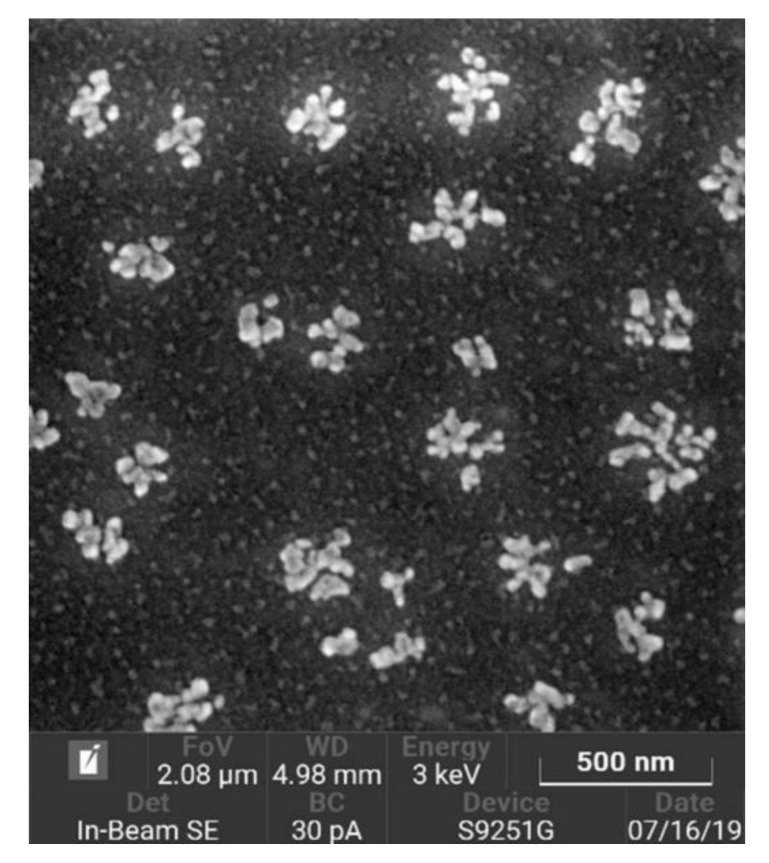
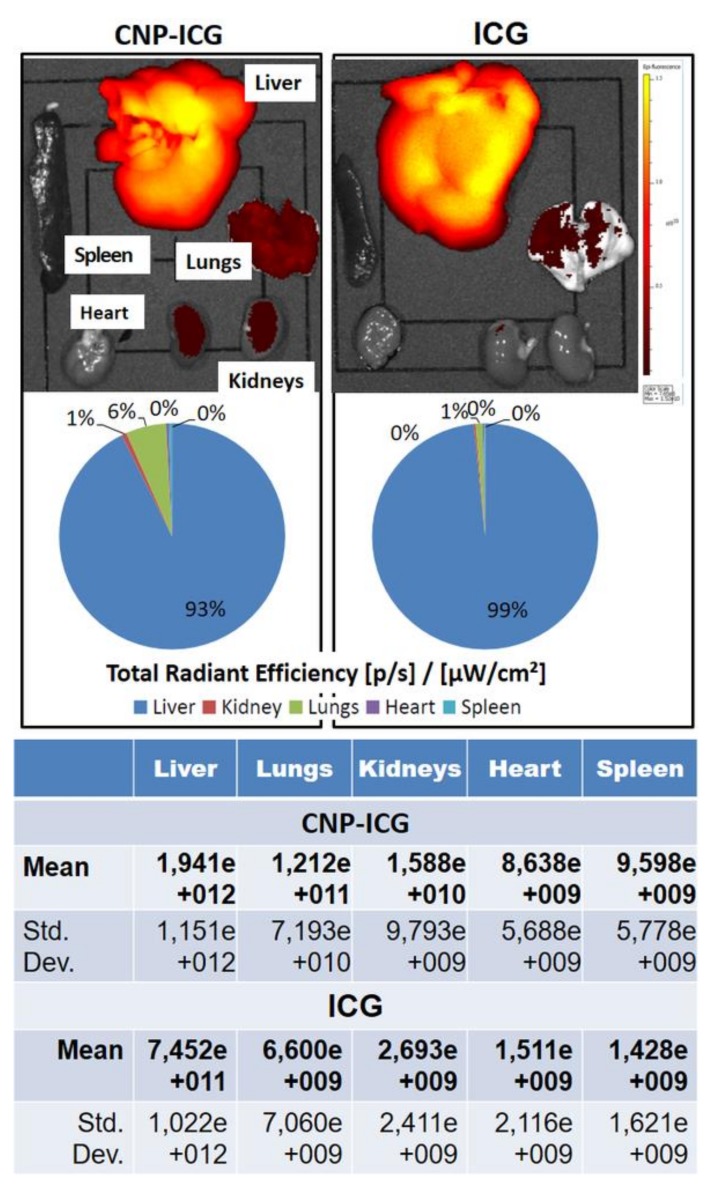
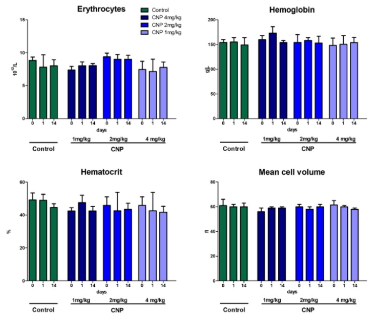
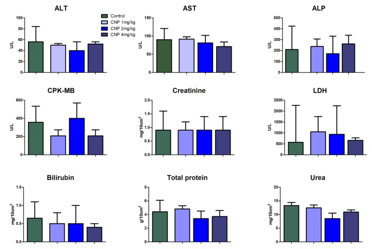
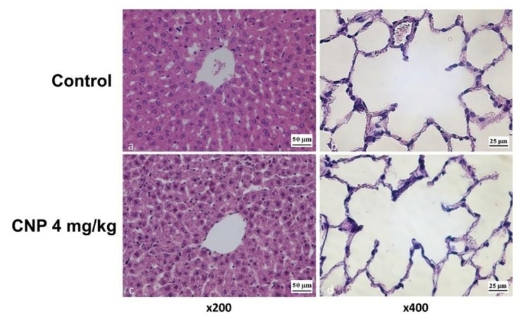
: The effect of unmodified chitosan nanoparticles with a size of ~100 nm and a weakly positive charge on blood coagulation, metabolic activity of cultured cardiomyocytes, general toxicity, biodistribution, and reactive changes in rat organs in response to their single intravenous administration at doses of 1, 2, and 4 mg/kg was studied. Chitosan nanoparticles (CNPs) have a small cytotoxic effect and have a weak antiplatelet and anticoagulant effect. Intravenous administration of CNPs does not cause significant hemodynamic changes, and 30 min after the CNPs administration, they mainly accumulate in the liver and lungs, without causing hemolysis and leukocytosis. The toxicity of chitosan nanoparticles was manifested in a dose-dependent short-term delay in weight gain with subsequent recovery, while in the 2-week observation period no signs of pain and distress were observed in rats. Granulomas found in the lungs and liver indicate slow biodegradation of chitosan nanoparticles. In general, the obtained results indicate a good tolerance of intravenous administration of an unmodified chitosan suspension in the studied dose range.

Keywords: biodistribution; blood compatibility; chitosan; in vivo treatment; polymer nanoparticles; safety evaluation.


Conflict of interest statement

The authors declare no conflict of interest.

Figures

Figure 1
Figure 1
Scanning electron microscopy (SEM) image of chitosan nanoparticles. Clusters of nanoparticles are not aggregates but individual particles that have come together during drying.
Figure 2
Figure 2
Absorbance (A) of rat plasma 60 min after intravenous administration of the suspension of chitosan nanoparticles (n = 7). +Control—positive control – artificially hemolyzed 5 ± 2% red blood cell suspension (n = 5); –Control—negative control (saline, n = 5). Mean ± SEM. * – p < 0.05 with respect to +Control. n.s.—nonsignificant. Increased plasma-free hemoglobin level is a sign of hemolysis. If intravenously administrated CNPs cause in vivo hemagglutination and hemolysis, then free hemoglobin should appear in the blood plasma samples, which is determined by the absorption values at a wavelength of 540 nm.
Figure 3
Figure 3
Platelet hemostasis in the ADP-induced platelet aggregation test with 0.12% CNP suspension added to human plasma. Mean ± standard error. In the ADP test, the CNPs significantly inhibited platelet aggregation in comparison with the control. * – p < 0.05 with respect to Control.
Figure 4
Figure 4
The effect of a suspension of chitosan nanoparticles on the viability of cultured cardiomyocytes. Cell viability was measured by MTT assay. Absorbance (A) positively correlates with number of alive cells. CNPs reduce cell viability in tested concentrations only after 72 h incubation. Mean ± SEM. * – p < 0.05 with respect to basal.
Figure 5
Figure 5
In loco biodistribution pattern of free and CNP-immobilized ICG 30 min after intravenous administration. The diagrams show the percentage of fluorophore accumulation in organs measured as total radiation efficiency ([p/s]/[µW/cm2]). Percentage are calculated from the mean of total radiant efficiency (given in the table) for each organ (n = 3).
Figure 6
Figure 6
Dynamics of body weight after intravenous administration of CNPs (% of the baseline). The Mann–Whitney test was used to assess the significance of differences between groups. Data shown are median ± range.
Figure 7
Figure 7
Mass ratios of rat organs 14 days after intravenous administration of CNP suspension. Median ± range. The ratio of organ mass to body weight is used in toxicology to detect target organs of the toxicant. There were no significant effects of CNPs on mean weight ratios of the organs.
Figure 8
Figure 8
Dynamics of rat hemoglobin and erythrocyte parameters at different time points (0, 1 and 14 days) after intravenous injection of CNPs (1–4 mg/kg). Data represent the median ± range. These data complement results of the analysis of the level of free hemoglobin in the blood plasma of rats after administration of CNPs. If CNPs caused significant intravascular hemolysis and thrombosis, all red cell mass parameters (erythrocytes, hematocrit, and hemoglobin) can be decreased by CNPs–erythrocyte interactions, while MCV within normal range in the first day after intravenous administration of CNPs. The results of hematological analysis did not reveal significant changes in the parameters of red blood.
Figure 9
Figure 9
Dynamics of rat leukocyte parameters. Median ± range. Changes in white blood cells count were observed 1 day after injection, which were caused by minor surgical intervention (venesection). An increase in the level of white blood cells in the blood is a sign of a systemic immune response to tissue/organ injury or invasion. In this study, the dose of chitosan nanoparticles administered intravenously was not enough to cause a significant increase in white blood cell levels.
Figure 10
Figure 10
Rat blood biochemical parameters at 14 days after intravenous CNP administration. Median ± range. CNPs were mostly accumulated in the liver and in a lesser extent in the lungs. Kidneys, spleen, and heart accumulated about one percent of all nanoparticles. CNP could affect these organs and impair their functions. Biochemical parameters related to the liver, kidney, and heart did not reveal any signs of injury or malfunction.
Figure 11
Figure 11
The histological pattern of rat organs at 14 days after a single injection of a CNP-4 suspension (4 mg/kg). Tissues were stained with hematoxylin/eosin.
Figure 12
Figure 12
Thickness (μm) of rat alveolar septa at 14 days after intravenous administration of CNP suspension. Mann–Whitney test. Median ± range.
Figure 13
Figure 13
The lungs and liver of rats at 14 days after CNP administration 4 mg/kg. Immunohistochemistry analysis with antibodies to CD68+. Areas with CD68+-macrophage granulomas.
Figure 14
Figure 14
Rat livers 2 h after administration of CNP suspension (16 mg/kg). Grocott’s staining technique: (a) hepatocytes of control group (100×), (b) the same hepatocytes at magnification 1000×; (c) CNP group, hepatic lobule, area of the central vein, hepatocytes with internalized CNPs (100×); (d) the same hepatocytes at magnification 1000×.
Figure 15
Figure 15
Rat lungs 2 h after administration of CNP suspension (16 mg/kg). Grocott’s staining technique revealed: (a) the alveoli of the lung of control rat (400×); (b) control rat lung artery (400×); (c) interstitial macrophages (arrows) containing CNPs in the alveolar septa (400×); (d) rat pulmonary artery and muscle layers with stained CNPs (400×).

References

    1. Kato Y., Onishi H., Machida Y. Contribution of chitosan and its derivatives to cancer chemotherapy. In Vivo. 2005;19:301–310. - PubMed
    1. Cheung R.C., Ng T.B., Wong J.H., Chan W.Y. Chitosan: An update on potential biomedical and pharmaceutical applications. Mar. Drugs. 2015;13:5156–5186. doi: 10.3390/md13085156. - DOI - PMC - PubMed
    1. Muxika A., Etxabide A., Uranga J., Guerrero P., de la Caba K. Chitosan as a bioactive polymer: Processing, properties and applications. Int. J. Biol. Macromol. 2017;105:1358–1368. doi: 10.1016/j.ijbiomac.2017.07.087. - DOI - PubMed
    1. Shariatinia Z. Pharmaceutical applications of chitosan. Adv. Colloid Interface Sci. 2019;263:131–194. doi: 10.1016/j.cis.2018.11.008. - DOI - PubMed
    1. Dubashynskaya N.V., Poshina D.N., Raik S.V., Urtti A., Skorik Y.A. Polysaccharides in ocular drug delivery. Pharmaceutics. 2020;12:22. doi: 10.3390/pharmaceutics12010022. - DOI - PMC - PubMed

LinkOut - more resources