Review
. 2020 Apr 23;21(8):2998.
doi: 10.3390/ijms21082998.

From Snapshots to Flipbook-Resolving the Dynamics of Ribosome Biogenesis with Chemical Probes

Affiliations
Review

From Snapshots to Flipbook-Resolving the Dynamics of Ribosome Biogenesis with Chemical Probes

Lisa Kofler et al. Int J Mol Sci. .

Abstract

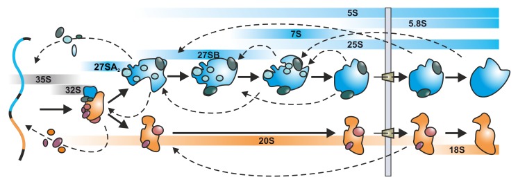
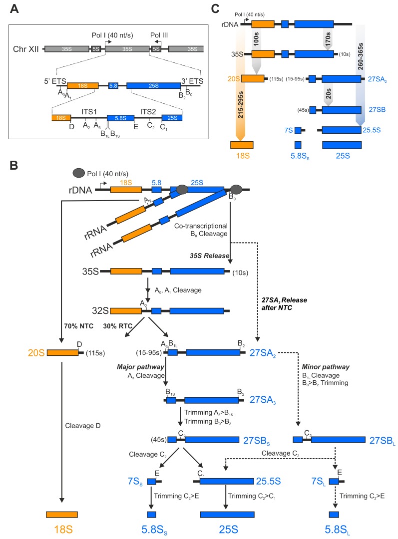
The synthesis of ribosomes is one of the central and most resource demanding processes in each living cell. As ribosome biogenesis is tightly linked with the regulation of the cell cycle, perturbation of ribosome formation can trigger severe diseases, including cancer. Eukaryotic ribosome biogenesis starts in the nucleolus with pre-rRNA transcription and the initial assembly steps, continues in the nucleoplasm and is finished in the cytoplasm. From start to end, this process is highly dynamic and finished within few minutes. Despite the tremendous progress made during the last decade, the coordination of the individual maturation steps is hard to unravel by a conventional methodology. In recent years small molecular compounds were identified that specifically block either rDNA transcription or distinct steps within the maturation pathway. As these inhibitors diffuse into the cell rapidly and block their target proteins within seconds, they represent excellent tools to investigate ribosome biogenesis. Here we review how the inhibitors affect ribosome biogenesis and discuss how these effects can be interpreted by taking the complex self-regulatory mechanisms of the pathway into account. With this we want to highlight the potential of low molecular weight inhibitors to approach the dynamic nature of the ribosome biogenesis pathway.

Keywords: chemical probing; feedback regulation; regulation of ribosome assembly; ribosome biogenesis; ribosome biogenesis inhibitors; ribosome trafficking.


Conflict of interest statement

The authors declare no conflict of interest.

Figures

Figure 1
Figure 1
Schematic overview of eukaryotic ribosome biogenesis in yeast. The synthesis of ribosomes starts in the nucleolus with the transcription of the 35S pre-rRNA, containing the 18S, 5.8S and 25S rRNAs and co-transcriptional protein assembly to form the small subunit processome. Upon endonucleolytic cleavage within the 35S pre-rRNA, the maturation pathways of the small and the large subunit are separated. The pre-40S subunit (orange) is rapidly exported to the cytoplasm, while the pre-60S subunit (blue) has to undergo numerous rearrangements and processing steps before its nuclear export. In the cytoplasm the last maturation and quality control steps occur before the subunits become competent for joining and translation. The respective (pre-)rRNAs are depicted in blue (rRNAs of the 60S subunit) and orange bars (rRNAs of the 40S subunit), respectively. Dashed arrows symbolize the complex disassembly, recycling and association cycles of assembly factors during ribosome biogenesis.
Figure 2
Figure 2
Eukaryotic rRNA processing in yeast. (A) Schematic depiction of the yeast rDNA locus on chromosome XII comprising 150 tandem repeats. Each repeat contains the sequence for the 5S rRNA which is transcribed by Pol III and the 35S pre-rRNA (comprising the 18S, 5.8S and 25S rRNA) which is synthesized by Pol I with a transcription rate of about 40 nucleotides per second in the opposite direction [20]. (B) Schematic overview of the 35S pre-rRNA processing cascade. During the exponential growth phase 70% of the nascent transcripts are co-transcriptionally cleaved at the A2 site (NTC: nascent transcript cleavage), while 30% are cleaved post-transcriptionally (RTC: released transcript cleavage) [20]. (C) Lifetimes (brackets) and processing times of pre-rRNAs in seconds (s). Koš and Tollervey (2010) determined the lifetimes and processing times of pre-RNAs via metabolic labeling [20]. The processing time of the 35S pre-rRNA to the indicated rRNAs is given in a range, since it is dependent on whether the A2 cleavage occurs co- or post-transcriptionally.
Figure 3
Figure 3
Impact of inhibitor treatment on eukaryotic ribosome biogenesis highlights the dynamics of the pathway. (A) The purification of the bait protein (light green protein) results in the copurification of a steady state particle population (indicated by curves) containing several distinct particles representing different maturation stages. (B) Upon short term inhibitor treatment (tshort) particles accumulate at the stage where the protein target is implicated in ribosome biogenesis, resulting in an entrapment of assembly factors that would be released after the block (magenta protein). This entrapment can cause a secondary blockage at earlier stages. Since downstream processes are not hindered at the time of drug application, these maturation steps still proceed for a certain period of time. Hence, assembly factors that leave the particle before the block (orange protein) will decrease in the particle population. However, also assembly factors that would bind downstream of the block (dark green protein) will decrease in the purification. Assembly factors that bind before the bait protein and would be released after the removal of the bait protein (blue protein) remain unchanged. (C) After long term treatment (tlong) a segregation of the particle population occurs, resulting in early population lacking factors (magenta factor) that are entrapped on the later population. Unassembled (orphan) proteins which cannot find their designated particle anymore (dark green factor) become degraded or precipitate. Dashed arrows symbolize the complex disassembly, recycling and association cycles of assembly factors during ribosome biogenesis. Bars represent particle-bound state of assembly factors (bound AF).
Figure 4
Figure 4
Diazaborine treatment allows tracing the ribosome biogenesis pathway from the nucleolar to the early cytoplasmic stage. While the steady state population of Nog1 in untreated cells (diazaborine t0) mainly contains 27SB pre-rRNA representing late nucleolar and early nucleoplasmic particles, treatment with diazaborine leads to a steady shift to a cytoplasmic population shortly after export. Since Nmd3 and Tif6 are the last two factors that leave the 60S-precursor before subunit joining, their cytoplasmic pool is significantly larger than the ones of Rlp24, Bud20 and Nog1, which dissociate shortly after export. As a consequence, significant amounts of Nmd3 and Tif6 can still be recycled shortly after drug application, while the recycling of Rlp24, Bud20 and Nog1 is blocked immediately (diazaborine t < 5 min). This fact allows tracking the particles that already contained Nog1 at the time of diazaborine application over time. The small nucleolar peak of Nog1 originates from de novo synthesis and import of Nog1, which is not affected by diazaborine and becomes detectable after about 15 minutes of drug treatment (diazaborine t >>5 min) [63].

References

    1. Baßler J., Hurt E. Eukaryotic Ribosome Assembly. Annu. Rev. Biochem. 2019;88:281–306. doi: 10.1146/annurev-biochem-013118-110817. - DOI - PubMed
    1. Klinge S., Woolford J.L. Ribosome assembly coming into focus. Nat. Rev. Mol. Cell Biol. 2019;20:116–131. doi: 10.1038/s41580-018-0078-y. - DOI - PMC - PubMed
    1. Bohnsack K.E., Bohnsack M.T. Uncovering the assembly pathway of human ribosomes and its emerging links to disease. EMBO J. 2019;38 doi: 10.15252/embj.2018100278. - DOI - PMC - PubMed
    1. Warner J.R. The economics of ribosome biosynthesis in yeast. Trends Biochem. Sci. 1999;24:437–440. doi: 10.1016/S0968-0004(99)01460-7. - DOI - PubMed
    1. Zaragoza D., Ghavidel A., Heitman J., Schultz M.C. Rapamycin Induces the G0 Program of Transcriptional Repression in Yeast by Interfering with the TOR Signaling Pathway. Mol. Cell Biol. 1998;18:4463–4470. doi: 10.1128/MCB.18.8.4463. - DOI - PMC - PubMed