Review
. 2021 Sep 1;11(9):a038372.
doi: 10.1101/cshperspect.a038372.

Structure and Function of Influenza Polymerase

Affiliations
Review

Structure and Function of Influenza Polymerase

Joanna M Wandzik et al. Cold Spring Harb Perspect Med. .

Abstract

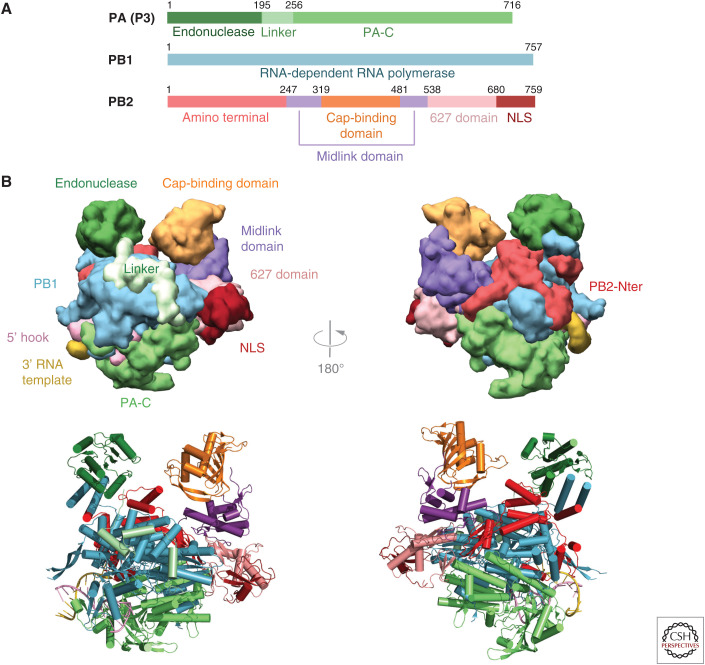
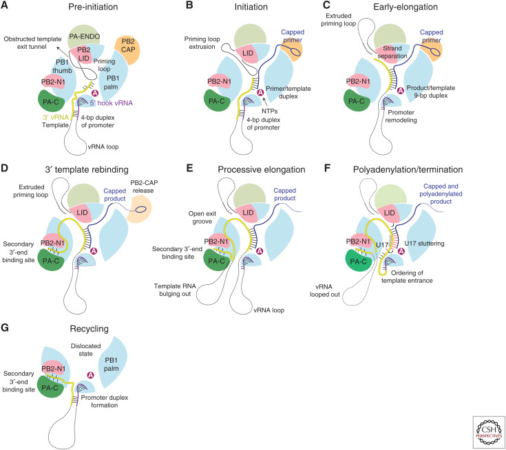
Influenza polymerase (FluPol) plays a key role in the viral infection cycle by transcribing and replicating the viral genome. FluPol is a multifunctional, heterotrimeric enzyme with cap-binding, endonuclease, RNA-dependent RNA polymerase and polyadenylation activities. It performs its functions in the context of the viral ribonucleoprotein particle (RNP), wherein the template viral RNA is coated by multiple copies of viral nucleoprotein. Moreover, it interacts with a number of host proteins that are essential cofactors and, consequently, adaptive mutations in the polymerase are required for crossing the avian-human species barrier. In this review, we show how mechanistic understanding of how FluPol performs its multiple functions has greatly advanced over the last decade through determination of high-resolution structures by X-ray crystallography and cryo-electron microscopy. These have revealed not only the detailed architecture of FluPol but highlighted the remarkably conformational flexibility that is inherent to its functioning as a dynamic RNA synthesis machine. Structural studies are also underpinning current attempts to develop next-generation anti-influenza drugs that directly target FluPol.


Figures

Figure 1.
Figure 1.
Overall structure of influenza polymerase. (A) Schematic of the three subunits showing major domains. Colors used throughout this review are PA-N endonuclease (dark green), PA linker (light green), PA-C (medium green), PB1 (cyan), PB2-N (red), PB2 midlink (purple), PB2 cap-binding domain (orange), PB2 627 domain (salmon), PB2 NLS domain (dark red). (B) Front and back views of influenza polymerase in the transcription active conformation in surface (top) and ribbon (bottom) representation showing the major domains colored as in A. Drawn from PDB: 4WSB.
Figure 2.
Figure 2.
Cap-snatching. (A) Capped RNA primer (slate blue) binding to the PB2 cap-binding and midlink domains and descending into the PB1 active site to initiation transcription. Drawn from PDB: 6QCX. (B) Close-up of (left) cap-analog m7GTP (slate blue) and (right) VX787/pimodivir (pink) binding in the influenza A cap-binding domain showing conserved interacting residues (orange sticks). Drawn from PDB: 4CB4 and 6EUV, respectively. (C) Surface representation of influenza A endonuclease with bound baloxavir acid (BXA) in the active site, showing two divalent cations (purple spheres) and conserved interacting residues (yellow sticks). The substitution I38T confers partial resistance to the drug. Drawn from PDB: 6FS6. (D) Schematic of how cap-snatching is performed. FluPol (gray surface with cap-binding domain in orange and endonuclease in green) binds to the serine 5 phosphorylated (pS5) carboxy-terminal domain (CTD) of host polymerase II (Pol II, light blue surface). This gives FluPol access to emerging nascent capped transcripts (red) that bind to the cap-binding domain and are cleaved 10–15 nt downstream by the endonuclease. The cap-binding domain then rotates to direct the capped primer into the PB1 RNA synthesis active site to initiate transcription.
Figure 3.
Figure 3.
Host–protein interacting domains. (A) Ribbon diagram showing the closed state of the PB2 627-NLS double domain. (B) Binding of the PB2 NLS domain, which carries a bipartite nuclear localization signal (NLS) in its carboxy-terminal extremity, to nuclear import factor importin-α. Drawn from PDB: 2JDQ. (C) Serine 5 phosphorylated Pol II CTD binding to influenza A, B, and C polymerases. In each case, two distinct sites are observed.
Figure 4.
Figure 4.
Promoter binding. (A) Schematic of the vRNA promoter structure that comprises the conserved 5′ (pink) and 3′ (yellow) extremities of each genomic vRNA segment. Nucleotides 1–10 of the 5′ end form a stem-loop structure called the “hook,” which binds tightly to the polymerase and acts as an allosteric regulator of polymerase function. Nucleotides 1–9 of the 3′ end are single-stranded and flexible. The distal duplex region is formed by interstrand base-pairs. The cRNA promoter forms a similar structure, but the single-stranded region of the 3′ end is 2 nt longer. (B) Mode A promoter binding showing the 5′ hook bound in a pocket between the PB1 and PA-C domains and the 3′ end of the template entering the PB1 RNA synthesis site. (C) Mode B promoter binding showing the vRNA 3′ end binding in the “secondary 3′-end binding site.”
Figure 5.
Figure 5.
Polymerase conformational flexibility. (A) Schematic showing the FluPol core, comprising PA-C, PB1, and PB2-N, to which are flexibly attached the peripheral PA endonuclease and PB2 midlink, cap, 627-, and NLS domains. (B) Surface representation of domain arrangement in the transcriptionally active state with bound full vRNA promoter. Drawn from PDB: 4WSB. (C) Domain arrangement in the transcriptionally inactive state of FluB polymerase with bound cRNA 5′ end only. Drawn from PDB: 5EPI. (D) Domain arrangement in the transcriptionally inactive state of FluC polymerase with no bound RNA (apo). Similar to C, but endonuclease and 627-domains orientated differently. Drawn from PDB: 5D9A. (E) Symmetric dimeric form of influenza A polymerase as observed in the crystal. The conformation of each monomer is as in D. Drawn from PDB: 6QNW.
Figure 6.
Figure 6.
PB1 RNA synthesis active site. (A) Ribbon diagram showing the conserved core polymerase architecture of the PB1 subunit comprising a right-handed arrangement of fingers (cyan), palm (red), and thumb (green). The fingertips loop (FT, blue) and priming loop (PL, magenta) occupy the central cavity where RNA synthesis occurs (see text). Additional PB1 specific features include the β-ribbon (orange), and the carboxy-terminal helices (wheat) that form a strong interface with PB2-N. (B) Mode A vRNA promoter binding to PB1 showing the template 3′ end, where RNA synthesis initiates, in the active site in close proximity to the tip of the priming loop (PL, magenta). Motif C (rose), a β-hairpin in the palm domain that carries two key metal binding aspartates and the two catalytic magnesium ions (gray spheres) are highlighted (see also Fig. 7). (C) Model of how the priming loop might promote vRNA to cRNA replication initiation by stabilizing the first phosphor-transfer reaction that requires ATP and GTP to assemble opposite U1 and C2 of the template, respectively.
Figure 7.
Figure 7.
Nucleotide addition cycle. (A) Details of the PB1 catalytic site showing key residues associated with the conserved polymerase motifs A–F and the two catalytic magnesium ions A and B (gray spheres). The primer/product RNA is in blue and the template strand in yellow. An incoming NTP, here a nonhydrolyzable UTP analog (UpNHpp), is shown in the precatalytic state. (B) First precatalytic step in the nucleotide addition cycle with incoming NTP stabilized by stacking on motif B Met409 (same structure as in A). (C) Postcatalytic, pretranslocation step in which the nucleotide has been added onto the product releasing pyrophosphate (PPi). (D) Translocation step, in which the template–product duplex has moved forward one position, bringing the next unpaired template base opposite the empty NTP binding site. Translocation may be assisted by flipping of the motif B loop into an alternative configuration.
Figure 8.
Figure 8.
Model for the complete transcription cycle (see text).

References

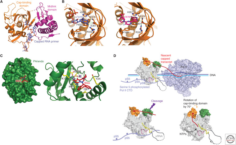
    1. Byrn RA, Jones SM, Bennett HB, Bral C, Clark MP, Jacobs MD, Kwong AD, Ledeboer MW, Leeman JR, McNeil CF, et al. 2015. Preclinical activity of VX-787, a first-in-class, orally bioavailable inhibitor of the influenza virus polymerase PB2 subunit. Antimicrob Agents Chemother 59: 1569–1582. 10.1128/AAC.04623-14 - DOI - PMC - PubMed
    1. Chang S, Sun D, Liang H, Wang J, Li J, Guo L, Wang X, Guan C, Boruah BM, Yuan L, et al. 2015. Cryo-EM structure of influenza virus RNA polymerase complex at 4.3 Å resolution. Mol Cell 57: 925–935. 10.1016/j.molcel.2014.12.031 - DOI - PubMed
    1. Chen KY, Santos Afonso ED, Enouf V, Isel C, Naffakh N. 2019. Influenza virus polymerase subunits co-evolve to ensure proper levels of dimerization of the heterotrimer. PLoS Pathog 15: e1008034. 10.1371/journal.ppat.1008034 - DOI - PMC - PubMed
    1. Chin AWH, Leong NKC, Nicholls JM, Poon LLM. 2017. Characterization of influenza A viruses with polymorphism in PB2 residues 701 and 702. Sci Rep 7: 11361. 10.1038/s41598-017-11625-y - DOI - PMC - PubMed
    1. Clark MP, Ledeboer MW, Davies I, Byrn RA, Jones SM, Perola E, Tsai A, Jacobs M, Nti-Addae K, Bandarage UK, et al. 2014. Discovery of a novel, first-in-class, orally bioavailable azaindole inhibitor (VX-787) of influenza PB2. J Med Chem 57: 6668–6678. 10.1021/jm5007275 - DOI - PubMed

Publication types