. 2020 May 12;117(19):10271-10277.
doi: 10.1073/pnas.2002888117. Epub 2020 Apr 27.

The structural basis for inhibition of ribosomal translocation by viomycin

Affiliations

The structural basis for inhibition of ribosomal translocation by viomycin

Ling Zhang et al. Proc Natl Acad Sci U S A. .

Abstract

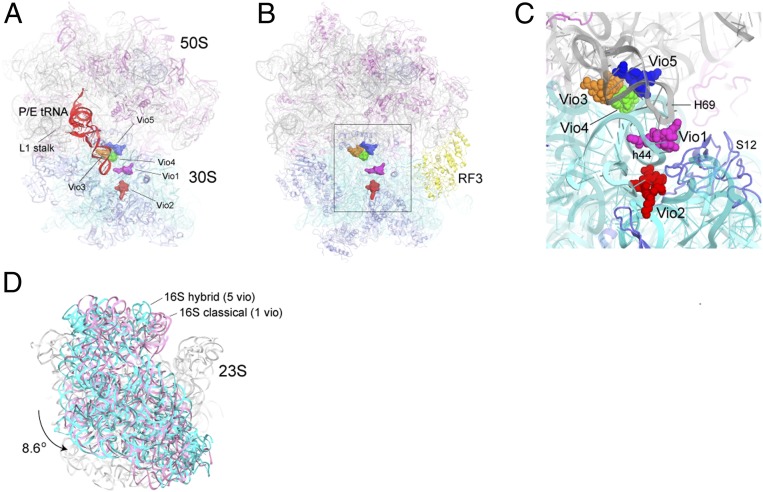
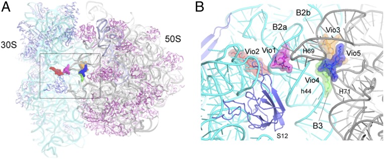
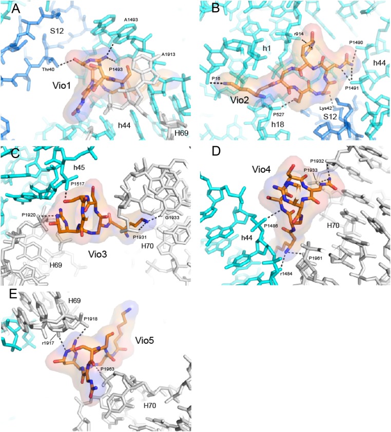
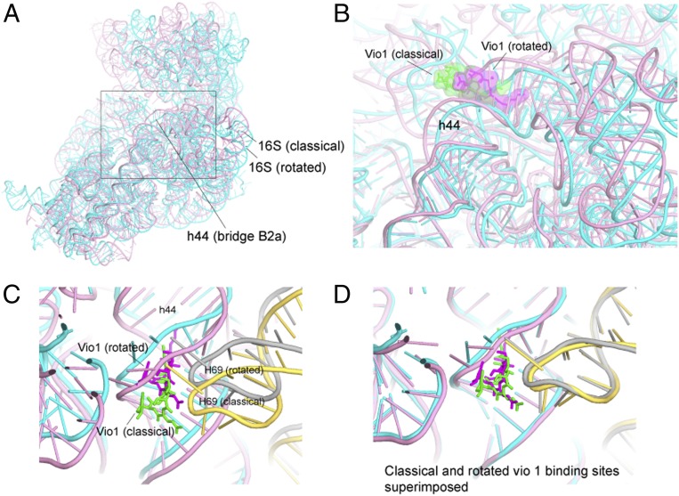
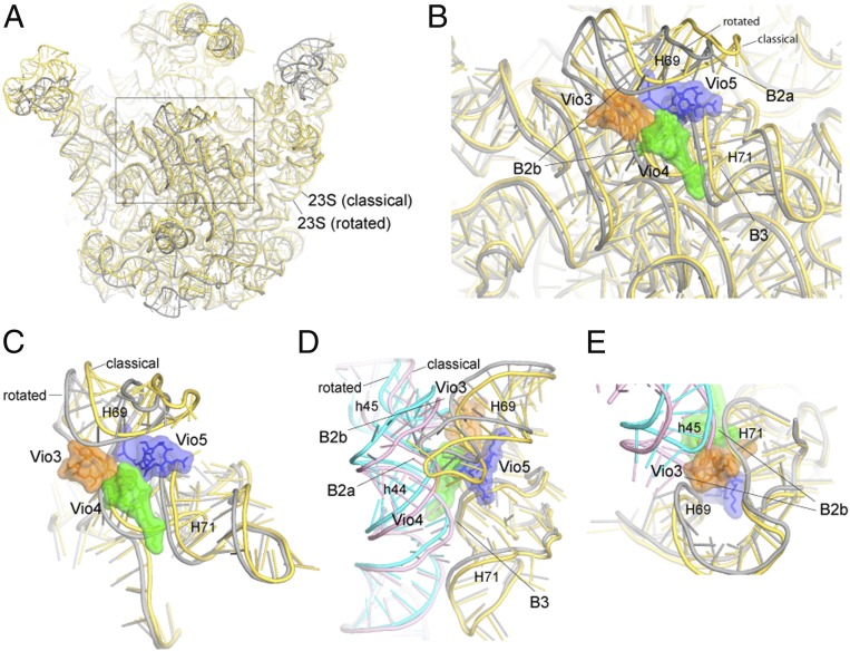
Viomycin, an antibiotic that has been used to fight tuberculosis infections, is believed to block the translocation step of protein synthesis by inhibiting ribosomal subunit dissociation and trapping the ribosome in an intermediate state of intersubunit rotation. The mechanism by which viomycin stabilizes this state remains unexplained. To address this, we have determined cryo-EM and X-ray crystal structures of Escherichia coli 70S ribosome complexes trapped in a rotated state by viomycin. The 3.8-Å resolution cryo-EM structure reveals a ribosome trapped in the hybrid state with 8.6° intersubunit rotation and 5.3° rotation of the 30S subunit head domain, bearing a single P/E state transfer RNA (tRNA). We identify five different binding sites for viomycin, four of which have not been previously described. To resolve the details of their binding interactions, we solved the 3.1-Å crystal structure of a viomycin-bound ribosome complex, revealing that all five viomycins bind to ribosomal RNA. One of these (Vio1) corresponds to the single viomycin that was previously identified in a complex with a nonrotated classical-state ribosome. Three of the newly observed binding sites (Vio3, Vio4, and Vio5) are clustered at intersubunit bridges, consistent with the ability of viomycin to inhibit subunit dissociation. We propose that one or more of these same three viomycins induce intersubunit rotation by selectively binding the rotated state of the ribosome at dynamic elements of 16S and 23S rRNA, thus, blocking conformational changes associated with molecular movements that are required for translocation.

Keywords: ribosome; translocation; viomycin.


Conflict of interest statement

The authors declare no competing interest.

Figures

Fig. 1.
Fig. 1.
Overall structures of viomycin-containing complexes. (A) Cryo-EM structure of the E. coli 70S ribosome in a complex with viomycin, mRNA, and a single P/E tRNA (red) in the hybrid state. The positions of the five bound viomycin molecules are indicated. (B and C) X-ray crystal structure of a viomycin-containing complex of the E. coli 70S ribosome bound with release factor RF3 (yellow), showing the positions of the five viomycin molecules and their surroundings in the ribosome, including 16S rRNA (cyan), 23S rRNA (gray), and ribosomal protein S12 (blue). (D) The 30S subunit in the cryo-EM structure shown in A (cyan) has undergone an 8.6° counterclockwise rotation relative to its orientation in the classical-state ribosome containing a single bound viomycin (pink) (21).
Fig. 2.
Fig. 2.
Positions of the viomycins relative to intersubunit bridges. (A) Overall view of the crystal structure of the 70S ribosome showing the positions of the five viomycins concentrated around the subunit interface. (B) Closeup view of the structure in A, showing the positions of the viomycins relative to structural features of the ribosome, including helices h44 of 16S rRNA and H69 and H71 of 23S rRNA and ribosomal protein S12. Three of the viomycins, Vio1, Vio3, and Vio4 bind at intersubunit bridges B2a, B2b, and B3.
Fig. 3.
Fig. 3.
Molecular Interactions between the viomycin and the ribosome. (A) Vio1 contacts helix h44 of 16S rRNA, H69 of 23S rRNA, and Thr40 of ribosomal protein S12 at intersubunit bridge B2a, similar to the single viomycin observed for the nonrotated ribosome complex (21). (B) Vio2 binds exclusively to the 30S subunit, contacting h1, h18, and h44 in 16S rRNA and Lys42 and 43 of protein S12. (C) Vio3 binds at intersubunit bridge B2b, contacting h45 in 16S rRNA and H69 and H70 of 23S rRNA. (D) Vio4 binds at intersubunit bridge B3, near the axis of intersubunit rotation, contacting h44 in 16S rRNA and H70 in 23S rRNA. (E) Vio5 binds between H69 and H70 of 23S rRNA, likely restricting movement of H69 during intersubunit rotation. The viomycin structures are shown in all-atom and transparent surface rendering. The 16S rRNA is shown in cyan, 23S rRNA in gray, and S12 in blue. RNA phosphate moieties are labeled by P, and riboses are labeled by r.
Fig. 4.
Fig. 4.
Vio1 binds to classical and rotated states. (A) Overall view of 16S rRNA in the classical (pink) and rotated (cyan) states as aligned on the core of 23S rRNA. (B) Movement of the Vio1 binding site between the classical and the rotated states. (C) H69 of 23S rRNA moves synchronously with h44 at the Vio1-binding site. (D) Alignment on 16S rRNA shows that the conformation of the Vio1-binding site is preserved upon intersubunit rotation. Vio1 is shown in green (classical) and magenta (rotated); 23S rRNA is shown in yellow (classical) and gray (rotated).
Fig. 5.
Fig. 5.
Binding of Vio3, Vio4, and Vio5 interferes with conformational changes associated with intersubunit rotation. (A) Superimposition of 23S rRNA in the classical (yellow) and rotated (gray) states. (B) Vio3, Vio4, and Vio5 cluster at the site of maximum movement of 23S rRNA components of intersubunit bridges B2b and B3. (C) Rotated view of B. (D) Rotational movement of 16S rRNA and compensating conformational changes in 23S rRNA. (E) Vio3 connects h45 and H69 to H70; in the classical state, h45 would create a steric clash with Vio3. In addition, both Vio3 and Vio5 (C) bridge H69 to H70, interfering with movement of H69.

References

    1. Akbergenov R., et al. , Molecular basis for the selectivity of antituberculosis compounds capreomycin and viomycin. Antimicrob. Agents Chemother. 55, 4712–4717 (2011). - PMC - PubMed
    1. Liou Y.-F., Tanaka N., Dual actions of viomycin on the ribosomal functions. Biochem. Biophys. Res. Commun. 71, 477–483 (1976). - PubMed
    1. Modolell J., Vázquez D., The inhibition of ribosomal translocation by viomycin. Eur. J. Biochem. 81, 491–497 (1977). - PubMed
    1. Polikanov Y. S., Aleksashin N. A., Beckert B., Wilson D. N., The mechanisms of action of ribosome-targeting peptide antibiotics. Front. Mol. Biosci. 5, 48 (2018). - PMC - PubMed
    1. Yamada T., Nierhaus K. H., Viomycin favours the formation of 70S ribosome couples. Mol. Gen. Genet. 161, 261–265 (1978). - PubMed

Publication types

MeSH terms