. 2020 Apr 27;11(4):294.
doi: 10.1038/s41419-020-2475-3.

The YY1/miR-548t-5p/CXCL11 signaling axis regulates cell proliferation and metastasis in human pancreatic cancer

Affiliations

The YY1/miR-548t-5p/CXCL11 signaling axis regulates cell proliferation and metastasis in human pancreatic cancer

Wan-Li Ge et al. Cell Death Dis. .

Expression of concern in

Abstract

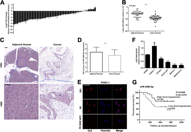
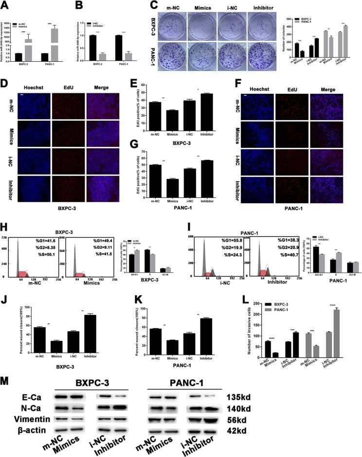
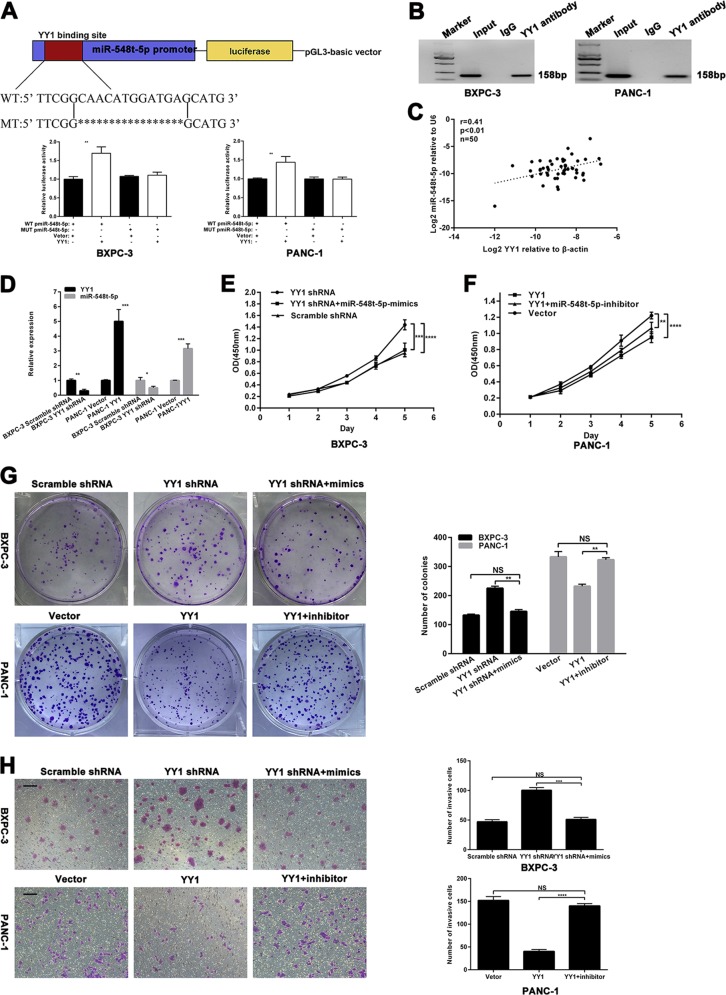
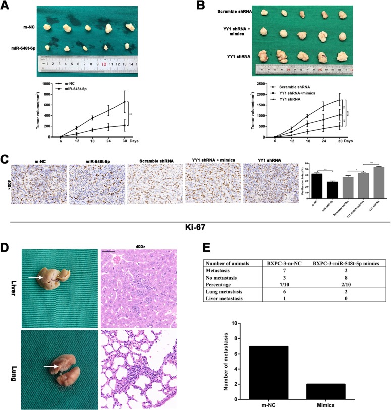
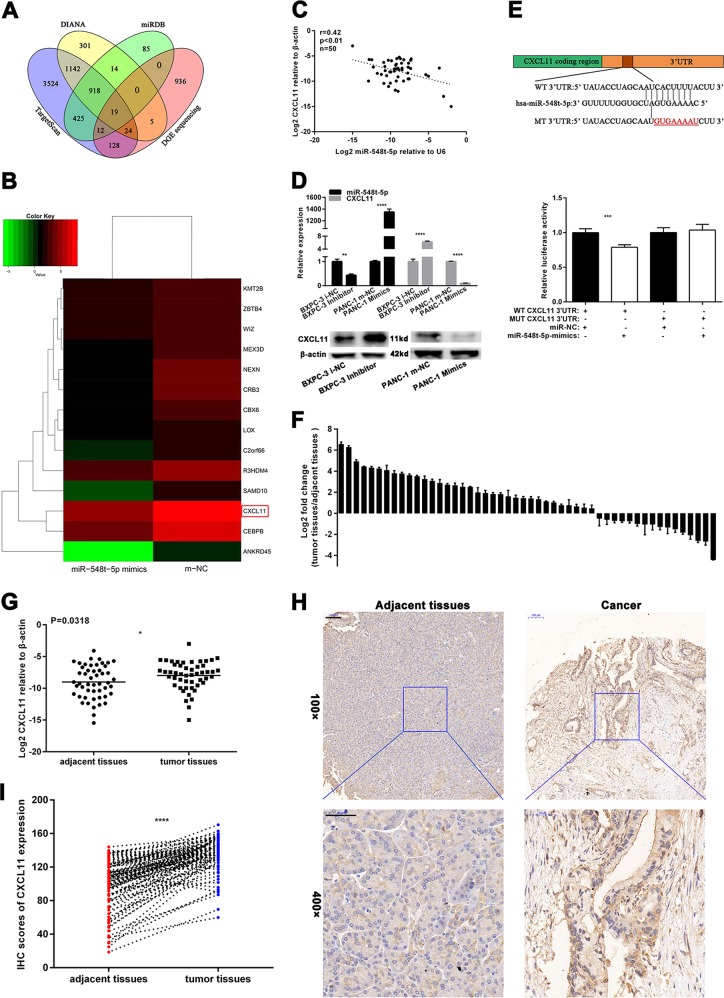
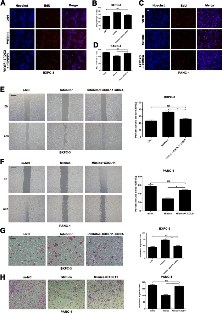
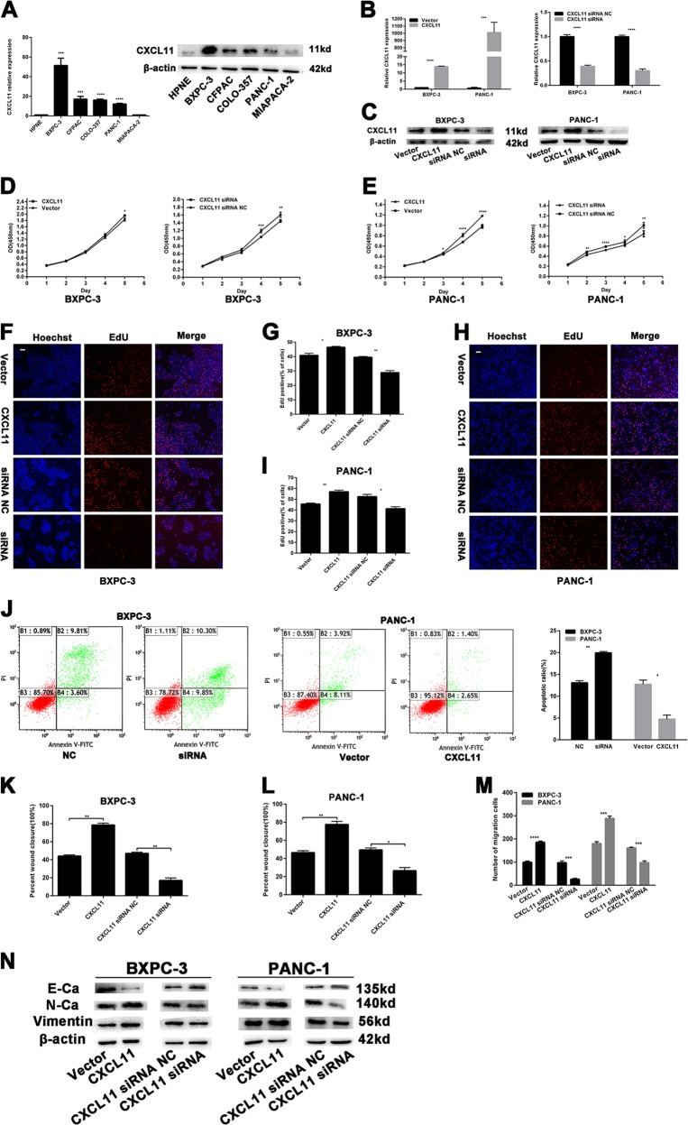
Pancreatic cancer (PC) is a malignant tumor with a poor prognosis and high mortality. However, the biological role of miR-548t-5p in PC has not been reported. In this study, we found that miR-548t-5p expression was significantly decreased in PC tissues compared with adjacent tissues, and that low miR-548t-5p expression was associated with malignant PC behavior. In addition, high miR-548t-5p expression inhibited the proliferation, migration, and invasion of PC cell lines. Regarding the molecular mechanism, the luciferase reporter gene, chromatin immunoprecipitation (ChIP), and functional recovery assays revealed that YY1 binds to the miR-548t-5p promoter and positively regulates the expression and function of miR-548t-5p. miR-548t-5p also directly regulates CXCL11 to inhibit its expression. A high level of CXCL11 was associated with worse Tumor Node Metastasis (TNM) staging in patients with PC, enhancing proliferation and metastasis in PC cells. Our study shows that the YY1/miR-548t-5p/CXCL11 axis plays an important role in PC and provides a new potential candidate for the treatment of PC.


Conflict of interest statement

The authors declare that they have no conflict of interest.

Figures

Fig. 1
Fig. 1. miR-548t-5p is downregulated in PC.
a qRT-PCR analysis of miR-548t-5p expression in 50 pairs of human PC tissues and adjacent non-neoplastic tissues. b As analyzed by qRT-PCR, miR-548t-5p expression in PC tissues was significantly lower than that in the corresponding adjacent non-neoplastic tissues. c Representative ISH images of miR-548t-5p expression. Scale bar, 100 μm in ×100, 50 μm in ×400. d A histogram showing the ISH results of miR-548t-5p expression in the 95 pairs of human PC tissues and adjacent non-neoplastic tissues. e MicroRNA FISH showed that miR-548t-5p was mostly located in the cytoplasm. Magnification, ×400; scale bar, 50 μm. f miR-548t-5p expression in normal human pancreatic ductal cell line hTERT‐HPNE and PC cell lines BXPC-3, CFPAC-1, COLO-357, MIAPACA-2, and PANC-1 by qRT-PCR. g Kaplan–Meier curves for overall survival (OS) by miR-548t-5p expression. The data are presented as mean ± SD from three independent experiments. *P ≤ 0.05, **P ≤ 0.01, ***P ≤ 0.001, ****P ≤ 0.0001.
Fig. 2
Fig. 2. The effects of miR-548t-5p on PC cells proliferation, migration, and invasion.
a, b Relative expression of miR-548t-5p by qRT-PCR in PC cells with miR-548t-5p-mimics and inhibitor. cg The colony formation assay (c) and EdU assay (dg) were performed to analyze the effects of miR-548t-5p-mimics or inhibitor on cell proliferation. Magnification, ×100; scale bar, 100 μm in d, f. h, i The flow cytometry assay analyzed the effect of miR-548t-5p on cell cycle status. j, k Wound-healing assays measured the effect of miR-548t-5p on PC cell migration ability. l Transwell assays measured the effect of miR-548t-5p on PC cell invasion ability. m EMT-related proteins were analyzed by western blotting assays in transfected PC cells. The data are presented as mean ± SD from three independent experiments. *P ≤ 0.05, **P ≤ 0.01, ***P ≤ 0.001, ****P ≤ 0.0001.
Fig. 3
Fig. 3. miR-548t-5p is regulated directly by the transcription factor YY1.
a Schematic diagram of the luciferase reporter construct containing the human miR-548t-5p promoter and the mutant YY1 construct containing miR-548t-5p promoter in which the presumed YY1-binding site was mutated. Results revealed that YY1 directly bound to the promoter region of has-miR-548t-5p. b ChIP assays were performed in BXPC-3-YY1-OE and PANC-1-YY1-OE cells. Lane 1, DNA marker; lane 2, input DNA; lane 3, DNA from cells immunoprecipitated with normal rabbit IgG; lane 4, DNA from cells immunoprecipitated with anti-YY1 antibody. c In human PC tissues, miR-548t-5p was positively correlated with YY1 expression. d YY1 and miR-548t-5p expression levels in YY1-overexpressing or YY1-knockdown cells were measured by qRT-PCR. eg CCK-8 and colony formation assays were performed to analyze proliferation in BXPC-3-YY1-knockdown cells transfected with miR-548t-5p-mimics and PANC-1-YY1-overexpression cells transfected with miR-548t-5p inhibitor. h Transwell assay was performed to analyze invasion in BXPC-3-YY1-knockdown cells transfected with miR-548t-5p-mimics and PANC-1-YY1-overexpression cells transfected with miR-548t-5p inhibitor. Magnification, ×100; scale bar, 100 μm. The data are presented as mean ± SD from three independent experiments. *P ≤ 0.05, **P ≤ 0.01, ***P ≤ 0.001, ****P ≤ 0.0001.
Fig. 4
Fig. 4. miR-548t-5p regulates the proliferation and metastasis of pancreatic cancer in vivo.
a Tumors were obtained from nude mice injected subcutaneously with BXPC-3 cells transfected with miR-548t-5p-mimics lentiviruses or control lentiviruses and the tumor volume was made into growth curve. b Tumors were obtained from nude mice injected subcutaneously with BXPC-3 cells transfected with YY1 shRNA lentiviruses, control lentiviruses, and YY1 shRNA lentiviruses in combination with miR-548t-5p-mimics lentiviruses and the tumor volume was made into growth curve. c IHC detection and quantification of Ki-67 protein expression in subcutaneous tumors from mice injected with BXPC-3 cells. Scale bar is 50 μm. d, e Representative pictures of lung and liver metastases are presented. The table listed the incidence of metastases in the nude mice treated with miR-548t-5p-mimics lentiviral vector or controls. Scale bar is 50 μm. The data are presented as mean ± SD from three independent experiments. *P ≤ 0.05, **P ≤ 0.01, ***P ≤ 0.001, ****P ≤ 0.0001.
Fig. 5
Fig. 5. miR-548t-5p directly targets CXCL11 in PC.
a The four-way Venn diagram indicates the numbers of genes that overlapped in three publicly available databases (DIANA, TargetScan, miRBD) and the DGE sequencing-based miR-548t-5p signature. b The heat map of the results of DGE sequencing based on 14 candidate genes that were downregulated in BXPC-3-miR-548t-5p-mimics cells. Red color represents an expression level above mean, green color represents an expression lower than the mean. c In human PC tissues, CXCL11 was positively correlated with miR-548t-5p expression. d miR-548t-5p and CXCL11 expression level in miR-548t-5p-mimics or miR-548t-5p inhibitor cells were measured by qRT-PCR. e Luciferase reporter assay was conducted to verify that miR-548t-5p directly bound to the 3′-UTR region of CXCL11. Luciferase activity was analyzed in cells cotransfected with miR-548t-5p-mimics or negative control with pGL3-CXCL11-WT or pGL3-CXCL11-MUT. f qRT-PCR analysis of CXCL11 expression in 50 pairs of human PC tissues and adjacent non-neoplastic tissues. g As analyzed by qRT-PCR, CXCL11 expression in PC tissues was significantly lower than that in the corresponding adjacent non-neoplastic tissues. h Representative IHC images of CXCL11 expression. Scale bar, 100 μm in ×100, 50 μm in ×400. i A histogram showing the IHC results of CXCL11 expression in the 95 pairs of human PC tissues and adjacent non-neoplastic tissues. The data are presented as mean ± SD from three independent experiments. *P ≤ 0.05, **P ≤ 0.01, ***P ≤ 0.001, ****P ≤ 0.0001.
Fig. 6
Fig. 6. miR-548t-5p suppresses cell proliferation and metastasis by targeting CXCL11.
ad EdU assay was performed to analyze proliferation in BXPC-3-miR-548t-5p inhibitor cells transfected with CXCL11 siRNA and PANC-1-miR-548t-5p-mimics cells transfected with CXCL11-overexpression vector. Magnification, ×100; scale bar, 100 μm in a and c. e, f Wound-healing assays were performed to analyze migration in BXPC-3-miR-548t-5p inhibitor cells transfected with CXCL11 siRNA and PANC-1-miR-548t-5p-mimics cells transfected with CXCL11-overexpression vector. Magnification, ×100; scale bar, 100 μm. g, h Transwell assays were performed to analyze migration in BXPC-3-miR-548t-5p inhibitor cells transfected with CXCL11 siRNA and PANC-1-miR-548t-5p-mimics cells transfected with CXCL11-overexpression vector. Magnification, ×100; scale bar, 100 μm. The data are presented as mean ± SD from three independent experiments. *P ≤ 0.05, **P ≤ 0.01, ***P ≤ 0.001, ****P ≤ 0.0001.
Fig. 7
Fig. 7. The effects of CXCL11 on PC cells proliferation and migration.
a Relative expression of CXCL11 by qRT-PCR and western blotting in PC cell lines. b, c Relative expression of CXCL11 by qRT-PCR and western blotting in PC cells with CXCL11-overexpression vector and siRNA. d, e The CCK-8 assays were performed to analyze the effects of CXCL11 on cell proliferation. fi EdU assays were performed to analyze the effects of CXCL11 on cell proliferation. Magnification, ×100; scale bar, 100 μm. j The flow cytometry analysis of the effect of CXCL11 expression alteration on cell apoptosis. k, l Wound-healing assays measured the effect CXCL11 on PC cell migration ability. m Transwell assays measured the effect CXCL11 on PC cell migration ability. n EMT-related protein was analyzed by western blotting assays in transfected PC cells. The data are presented as mean ± SD from three independent experiments. *P ≤ 0.05, **P ≤ 0.01, ***P ≤ 0.001, ****P ≤ 0.0001.

References

    1. Siegel RL, Miller KD, Jemal A. Cancer statistics, 2019. CA Cancer J. Clin. 2019;69:7–34. doi: 10.3322/caac.21551. - DOI - PubMed
    1. Dal Molin M, et al. Very long-term survival following resection for pancreatic cancer is not explained by commonly mutated genes: results of whole-exome sequencing analysis. Clin. Cancer Res. 2015;21:1944–1950. doi: 10.1158/1078-0432.CCR-14-2600. - DOI - PMC - PubMed
    1. Grasso C, Jansen G, Giovannetti E. Drug resistance in pancreatic cancer: Impact of altered energy metabolism. Crit. Rev. Oncol. Hematol. 2017;114:139–152. doi: 10.1016/j.critrevonc.2017.03.026. - DOI - PubMed
    1. Griffin JF, Poruk KE, Wolfgang CL. Pancreatic cancer surgery: past, present, and future. Chin. J. Cancer Res. 2015;27:332–348. - PMC - PubMed
    1. Chin V, et al. Chemotherapy and radiotherapy for advanced pancreatic cancer. Cochrane Database Syst. Rev. 2018;3:CD011044. - PMC - PubMed

Publication types

Substances