. 2020 Apr 27;10(1):7047.
doi: 10.1038/s41598-020-63766-2.

Innate Immune Functions of Astrocytes are Dependent Upon Tumor Necrosis Factor-Alpha

Affiliations

Innate Immune Functions of Astrocytes are Dependent Upon Tumor Necrosis Factor-Alpha

Kyla R Rodgers et al. Sci Rep. .

Abstract

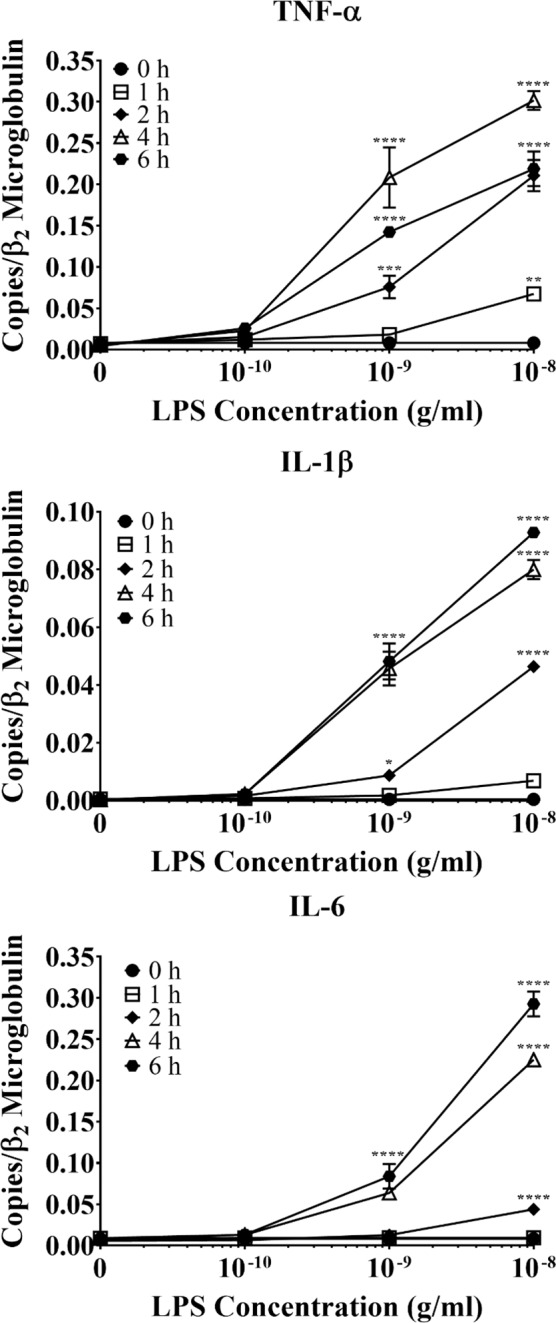
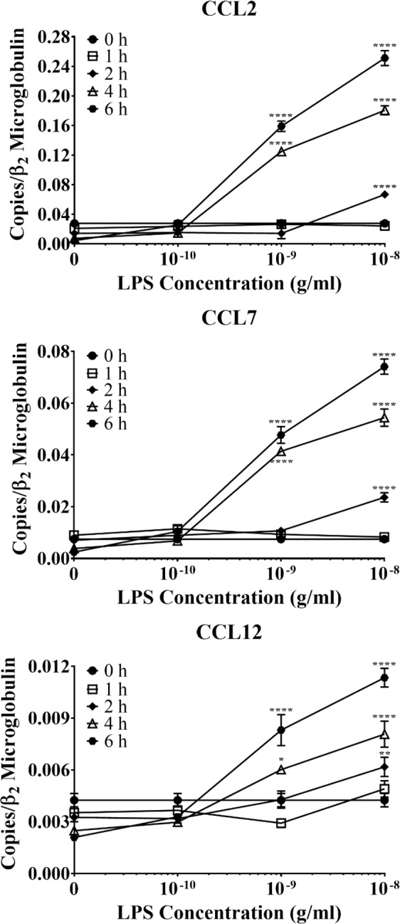
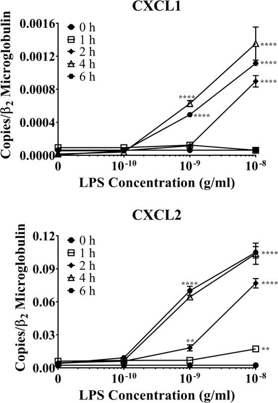
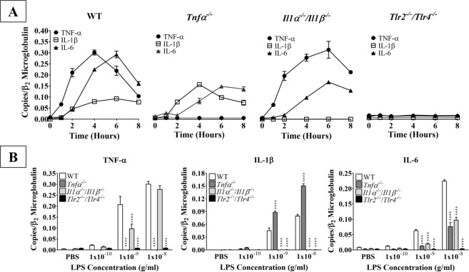
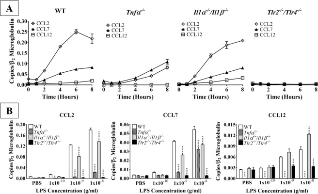
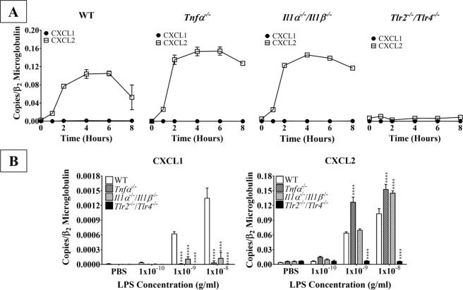
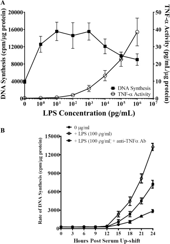
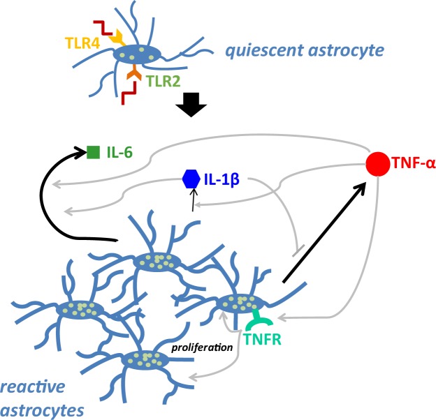
Acute inflammation is a key feature of innate immunity that initiates clearance and repair in infected or damaged tissues. Alternatively, chronic inflammation is implicated in numerous disease processes. The contribution of neuroinflammation to the pathogenesis of neurological conditions, including infection, traumatic brain injury, and neurodegenerative diseases, has become increasingly evident. Potential drivers of such neuroinflammation include toll-like receptors (TLRs). TLRs confer a wide array of functions on different cell types in the central nervous system (CNS). Importantly, how TLR activation affects astrocyte functioning is unclear. In the present study, we examined the role of TLR2/4 signaling on various astrocyte functions (i.e., proliferation, pro-inflammatory mediator production, regulatory mechanisms, etc) by stimulating astrocytes with potent exogenous TLR2/4 agonist, bacterial lipopolysaccharide (LPS). Newborn astrocytes were derived from WT, Tnfα-/-, Il1α-/-/Il1β-/-, and Tlr2-/-/Tlr4-/- mice as well as Sprague Dawley rats for all in vitro studies. LPS activated mRNA expression of different pro-inflammatory cytokines and chemokines in time- and concentration-dependent manners, and upregulated the proliferation of astrocytes based on increased 3H-thymidine update. Following LPS-mediated TLR2/4 activation, TNF-α and IL-1β self-regulated and modulated the expression of pro-inflammatory cytokines and chemokines. Polyclonal antibodies against TNF-α suppressed TLR2/4-mediated upregulation of astrocyte proliferation, supporting an autocrine/paracrine role of TNF-α on astrocyte proliferation. Astrocytes perform classical innate immune functions, which contradict the current paradigm that microglia are the main immune effector cells of the CNS. TNF-α plays a pivotal role in the LPS-upregulated astrocyte activation and proliferation, supporting their critical roles in in CNS pathogenesis.


Conflict of interest statement

The authors declare no competing interests.

Figures

Figure 1
Figure 1
The effect of LPS on primary WT astrocyte production of pro-inflammatory cytokines. Purified and subcultured murine WT astrocytes were incubated with graded concentrations of freshly prepared bacterial LPS or vehicle control (PBS). Cultures were terminated at 0, 1, 2, 4, or 6 h after stimulation. Total RNA was then extracted, and mRNA expression of pro-inflammatory cytokines was determined by qPCR. *p < 0.05; **p < 0.01; ***p < 0.001; ****p < 0.0001 (n = 3, results show one representative experiment out of 4).
Figure 2
Figure 2
The effect of LPS on primary WT astrocyte production of CCR2 ligands. Purified and subcultured murine WT astrocytes were incubated with graded concentrations of freshly prepared bacterial LPS or vehicle control (PBS). Cultures were terminated at 0, 1, 2, 4, or 6 h after stimulation. Total RNA was then extracted, and mRNA expression of CCR2 ligands was determined by qPCR. *p < 0.05; **p < 0.01; ***p < 0.001; ****p < 0.0001 (n = 3, results show one representative experiment out of 4).
Figure 3
Figure 3
The effect of LPS on primary WT astrocyte production of neutrophil-active chemokines. Purified and subcultured murine WT astrocytes were incubated with graded concentrations of freshly prepared bacterial LPS or vehicle control (PBS). Cultures were terminated at 0, 1, 2, 4, or 6 h after stimulation. Total RNA was then extracted, and mRNA expression of neutrophil-active chemokines was determined by qPCR. *p < 0.05; **p < 0.01; ***p < 0.001; ****p < 0.0001 (n = 3, results show one representative experiment out of 4).
Figure 4
Figure 4
Kinetics of pro-inflammatory cytokine production by LPS-stimulated WT and knockout astrocytes. (A) Purified and subcultured murine WT, Tnfα−/−, Il1α−/−/Il1β−/−, and Tlr2−/−/Tlr4−/− astrocytes were incubated with graded concentrations of freshly prepared bacterial LPS (0–10 ng/mL). Cultures were terminated at 0, 1, 2, 4, 6, or 8 h after stimulation. Total RNA was then extracted, and mRNA expression of TNF-α, IL-1β, and IL-6 was determined by qPCR (n = 3, results show one representative experiment out of 4). (B) Purified and subcultured murine WT, Tnfα−/−, Il1α−/−/Il1β−/−, and Tlr2−/−/Tlr4−/− astrocytes were incubated with graded concentrations of freshly prepared bacterial LPS (0–10 ng/mL). Cultures were terminated 4 h after stimulation. Total RNA was then extracted, and mRNA expression of TNF-α, IL-1β, and IL-6 was determined by qPCR. *p < 0.05; **p < 0.01; ***p < 0.001; ****p < 0.0001 (n = 3, results show one representative experiment out of 4).
Figure 5
Figure 5
Kinetics of CCR2 ligand production by LPS-stimulated WT and knockout astrocytes. (A) Purified and subcultured murine WT, Tnfα−/−, Il1α−/−/Il1β−/−, and Tlr2−/−/Tlr4−/− astrocytes were incubated with graded concentrations of freshly prepared bacterial LPS (0–10 ng/mL). Cultures were terminated at 0, 1, 2, 4, 6, or 8 h after stimulation. Total RNA was then extracted, and mRNA expression of CCL2, CCL7, and CCL12 was determined by qPCR. (B) Purified and subcultured murine WT, Tnfα−/−, Il1α−/−/Il1β−/−, and Tlr2−/−/Tlr4−/− astrocytes were incubated with graded concentrations of freshly prepared bacterial LPS (0–10 ng/mL). Cultures were terminated 4 h after stimulation. Total RNA was then extracted, and mRNA expression of CCL2, CCL7, and CCL12 was determined by qPCR. *p < 0.05; **p < 0.01; ***p < 0.001; ****p < 0.0001 (n = 3, results show one representative experiment out of 4).
Figure 6
Figure 6
Kinetics of neutrophil chemokine production by LPS-stimulated WT and knockout astrocytes. (A) Purified and subcultured murine WT, Tnfα−/−, Il1α−/−/Il1β−/−, and Tlr2−/−/Tlr4−/− astrocytes were incubated with graded concentrations of freshly prepared bacterial LPS (0–10 ng/mL). Cultures were terminated at 0, 1, 2, 4, 6, or 8 h after stimulation. Total RNA was then extracted, and mRNA expression of CXCL1 and CXCL2 was determined by qPCR. (B) Purified and subcultured murine WT, Tnfα−/−, Il1α−/−/Il1β−/−, and Tlr2−/−/Tlr4−/− astrocytes were incubated with graded concentrations of freshly prepared bacterial LPS (0–10 ng/mL). Cultures were terminated 4 h after stimulation. Total RNA was then extracted, and mRNA expression of CXCL1 and CXCL2 was determined by qPCR. *p < 0.05; **p < 0.01; ***p < 0.001; ****p < 0.0001 (n = 3 results show one representative experiment out of 4).
Figure 7
Figure 7
(A) Production of bioactive TNF-α by serum-deprived astrocytes as they re-enter cell cycle following LPS stimulation. Newborn rat astrocytes were rendered into G0 phase by serum deprivation and then allowed to re-enter the cell cycle via serum up-shift in the presence of graded concentrations of LPS. After a 24-h incubation, cell-free medium was collected to measure bioactive TNFα production and the rate of DNA synthesis using the WEHI and 3H-thymidine incorporation assays, respectively (n = 7–9 results show an average of three experiments). (B) LPS-stimulated cell cycle re-entry in astrocytes is TNF-α-dependent. Newborn astrocytes were rendered into G0 phase by serum deprivation and then allowed to re-enter the cell cycle via serum up-shift in the presence of freshly prepared LPS (100 pg/ml) with or without anti-TNF-α antibodies (10 U/mL). The 3H-thymidine incorporation assay was performed to measure rate of DNA synthesis. WT vs. LPS treatment: ***p < 0.05; **p < 0.001; *p < 0.0001. LPS treatment group vs. LPS + anti-TNF-α antibodies: !p < 0.001; !!p < 0.01; !!!p < 0.05 (n = 7–9 results show an average of three experiments).
Figure 8
Figure 8
The self-regulatory mechanisms underlying pro-inflammatory cytokine production following TLR2/4 activation. Feedback pathways were determined by quantifying mRNA production of each pro-inflammatory cytokine following stimulation with bacterial LPS. Arrow thickness indicates the relative magnitude of peak production. Black arrows indicate direct production of mediators by astrocytes; gray arrows indicate mechanisms of self-regulation, as determined by the effect of various knockouts on cytokine expression. Arrows with pointed heads indicate positive regulation, while arrows with flat, perpendicular lines indicate negative regulation. White dots on astrocytes represent GFAP expression.

References

    1. Verkhratsky A, Nedergaard M. Physiology of astroglia. Physiol. Rev. 2008;98:239–389. doi: 10.1152/physrev.00042.2016. - DOI - PMC - PubMed
    1. Ransom B, Behar T, Nedergaard M. New roles for astrocytes (stars at last) Trends Neurosci. 2009;26:520–522. doi: 10.1016/j.tins.2003.08.006. - DOI - PubMed
    1. Lee CY, Pappas GD, Kriho V, Huang BM, Yang HY. Proliferation of a subpopulation of reactive astrocytes following needle-insertion lesion in rat. Neurol. Res. 2003;25:767–776. doi: 10.1179/016164103101202156. - DOI - PubMed
    1. Pekny M, Pekna M. Astrocyte reactivity and reactive astrogliosis: costs and benefits. Physiol. Rev. 2014;94:1077–1098. doi: 10.1152/physrev.00041.2013. - DOI - PubMed
    1. Hennessy EJ, Parker AE, O’Neill LA. Targeting Toll-like receptors: emerging therapeutics? Nat. Rev. Drug Discov. 2010;9:293–307. doi: 10.1038/nrd3203. - DOI - PubMed

LinkOut - more resources