. 2020 May;27(5):450-460.
doi: 10.1038/s41594-020-0418-4. Epub 2020 Apr 27.

Reconstitution of translesion synthesis reveals a mechanism of eukaryotic DNA replication restart

Affiliations

Reconstitution of translesion synthesis reveals a mechanism of eukaryotic DNA replication restart

Thomas A Guilliam et al. Nat Struct Mol Biol. 2020 May.

Abstract

Leading-strand template aberrations cause helicase-polymerase uncoupling and impede replication fork progression, but the details of how uncoupled forks are restarted remain uncertain. Using purified proteins from Saccharomyces cerevisiae, we have reconstituted translesion synthesis (TLS)-mediated restart of a eukaryotic replisome following collision with a cyclobutane pyrimidine dimer. We find that TLS functions 'on the fly' to promote resumption of rapid replication fork rates, despite lesion bypass occurring uncoupled from the Cdc45-MCM-GINS (CMG) helicase. Surprisingly, the main lagging-strand polymerase, Pol δ, binds the leading strand upon uncoupling and inhibits TLS. Pol δ is also crucial for efficient recoupling of leading-strand synthesis to CMG following lesion bypass. Proliferating cell nuclear antigen monoubiquitination positively regulates TLS to overcome Pol δ inhibition. We reveal that these mechanisms of negative and positive regulation also operate on the lagging strand. Our observations have implications for both fork restart and the division of labor during leading-strand synthesis generally.


Conflict of interest statement

Competing Interests Statement

The authors declare no competing interests.

Figures

Extended Data Fig. 1
Extended Data Fig. 1. Pol η promotes TLS of lagging and leading-strand CPDs.
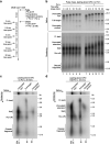
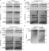
a, Purified Okazaki fragment processing and TLS proteins. b, Long exposure of the denaturing gel shown in Fig. 1b showing the diffuse ~1.7 kb stall product produced on the lagging-strand CPD template. c, Two-dimensional gel of the reaction performed in the absence of Pol η on the undamaged leading-strand template, shown in lane 1 of main text Fig. 1d. d, Two-dimensional gel of the reaction performed in the absence of Pol η on the leading-strand CPD template, shown in lane 7 of main text Fig. 1d. e, Two-dimensional gel of the reaction performed in the presence of 16 nM Pol η on the leading-strand CPD template, shown in lane 12 of main text Fig. 1d.
Extended Data Fig. 2
Extended Data Fig. 2. Leading-strand TLS occurs uncoupled from CMG.
a, Oligonucleotide competition assay performed in the absence or presence of Pol η. Reaction products were cleaved with SwaI to truncate stall products before resolution on a urea polyacrylamide gel. Addition of Pol η promotes extension of the stall product in the gap left behind from oligonucleotide-mediated recoupling. b, Reaction scheme for the pulse-chase experiment shown in (c). c, Pulse chase experiment on the leading-strand CPD template with 5 nM Pol η added 3 min into the pulse, at the start of the chase, or 10 min into the chase.
Extended Data Fig. 3
Extended Data Fig. 3. Pol δ, but not Pol ε, inhibits lagging and leading-strand TLS.
a, Pol δ titration into standard replication reactions on the undamaged leading-strand template containing Fen1 and Ligase. b, Denaturing gel of the reaction products from main text Fig. 3a. c, Standard replication reaction on the lagging-strand CPD template in the presence of 5 nM Pol η and increasing concentrations of Pol δ, as performed in Fig. 3a, but in the absence of Fen1 and Ligase. d, Pol δ titration into standard replication reactions on the undamaged leading-strand template. e, Reaction scheme for the pulse-chase experiment shown in (f). f, Pulse chase experiment on the leading-strand CPD template with 5 nM Pol η alone, or with 5 nM extra Pol ε, or Pol δ, added at the start of the chase.
Extended Data Fig. 4
Extended Data Fig. 4. Uncoupled replication forks display a recoupling defect in the absence of Pol δ.
a, Reaction scheme for the pulse-chase experiment shown in (b). b, Pulse chase experiment on the leading-strand CPD template in the absence of Pol δ and the absence or presence of 5 nM Pol η, added at the start of the chase. c, Two-dimensional gel of the 20 min time point shown in lane 6 of (b). d, Two-dimensional gel of the 20 min time point shown in lane 12 of (b).
Extended Data Fig. 5
Extended Data Fig. 5. PCNA monoubiquitination stimulates on the fly TLS.
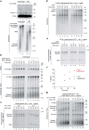
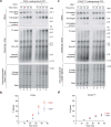
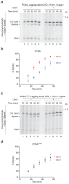
a, Western blot of PCNA from standard 60 min replication reactions on the leading-strand CPD template, or undamaged equivalent, in the absence or presence of Fen1 and Ligase. All reactions contained ubiquitin, Uba1, and Rad6–Rad18 in addition to standard replication proteins. Denaturing gel of reaction products is shown below. b, Standard replication reaction time course on the undamaged template performed in the absence or presence of Rad6–Rad18, Uba1, and ubiquitin. c, Standard replication reaction time course on the leading-strand CPD template in the presence of 2.5 nM Pol η and 0.3 nM or 2.5 nM Pol δ. Samples were treated with BamHI and SwaI to generate bypass and stall products prior to resolution on the urea polyacrylamide gel. d, Denaturing gel of the reaction products from Fig. 5c. e, Replication reaction time course performed on the leading-strand CPD template in the absence or presence of Uba1 or Rad6–Rad18. Reactions contained 2.5 nM Pol η, 2.5 nM Pol δ, 1 μM ubiquitin, 5 nM Fen1, and 5 nM Ligase, in addition to standard replication proteins. Urea polyacrylamide gel samples were treated with BamHI and SwaI to generate quantifiable bypass and stall products. f, Quantification of the data in (e) showing the percentage of bypass in the absence or presence of uba1 or Rad6–18. g, Replication reaction time course performed on the leading-strand CPD template in the presence of PCNA monoubiquitination machinery (Rad6–Rad18, Uba1, and ubiquitin) and Pol η and the absence or presence of Pol δ.
Extended Data Fig. 6
Extended Data Fig. 6. Characterization of PCNAK164R.
a, Standard replication reaction time course performed on the undamaged template with either wild type PCNA or PCNAK164R. b, Standard replication reactions on the leading-strand CPD template containing increasing amounts of wild type PCNA or PCNAK164R. Reactions contained 5 nM Pol η. c, Standard replication reactions on the leading-strand CPD template in the absence or presence of 2.5 nM Pol δ and increasing amounts of wild type PCNA. Reactions contained 5 nM Pol η. d, Standard replication reaction time course on the undamaged leading-strand template with either wild type PCNA or PCNAK164R in the presence of Fen1 and Ligase.
Extended Data Fig. 7
Extended Data Fig. 7. PCNA monoubiquitination stimulates on the fly TLS in the absence of Fen1 and Ligase.
a, Standard replication reaction time course performed with wild-type PCNA in the absence or presence of Uba1. Reactions contained 10 nM Pol η, 10 nM Pol δ, 250 nM ubiquitin, and 200 nM Rad6–Rad18, in addition to standard replication proteins. Urea polyacrylamide gel samples were treated with BamHI and SwaI to generate quantifiable bypass and stall products. b, Quantification of the percentage of bypass in the absence or presence of Uba1 as performed in (b). Data are plotted as the means and s.e.m. of three independent experiments. c, Standard replication reaction time course performed with PCNAK164R in the absence or presence of Uba1. Reactions contained 10 nM Pol η, 10 nM Pol δ, 250 nM ubiquitin, and 200 nM Rad6–Rad18, in addition to standard replication proteins. Urea polyacrylamide gel samples were treated with BamHI and SwaI to generate quantifiable bypass and stall products. d, Quantification of the percentage of bypass in the absence or presence of Uba1 as performed in (d). Data are plotted as the means and s.e.m. of three independent experiments.
Extended Data Fig. 8
Extended Data Fig. 8. PCNA monoubiquitination promotes lagging-strand TLS.
a, Standard replication reaction time course on the lagging-strand CPD template in the presence of 2.5 nM Pol η and 0.3125 nM or 2.5 nM Pol δ. Samples were treated with BamHI and SwaI to generate bypass and stall products prior to resolution on the urea polyacrylamide gel. b, Denaturing gel of the reaction products from Fig. 6a.
Fig. 1
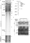
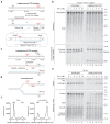
Fig. 1. Reconstitution of lagging- and leading-strand TLS.
(a) Schematic of the 9.7 kb ARS306 lagging-strand CPD template. The origin of replication (Ori), CPD site, and leading- (red) and lagging-strand (blue) replication products produced in the presence of Fen1 and Ligase are illustrated. A dashed line indicates lesion bypass and gap filling. Below is shown the position of the BamHI and SwaI restriction sites used for urea polyacrylamide gel analysis, in addition to the stall (165 nt) and bypass (187 nt) products generated by cleavage. (b) Standard replication assay on the lagging-strand CPD template and undamaged equivalent in the presence of Fen1 and Ligase (Cdc9) with increasing concentrations of Pol η. Urea polyacrylamide gel samples were treated with BamHI and SwaI to generate the stall and bypass products shown in (A) (used where indicated in subsequent figures). The ‘cut lead’ product was generated by cleavage of the leading strand by BamHI and SwaI. (c) Schematic of the 9.7 kb ARS306 leading-strand CPD template. Leading- (red) and lagging-strand (blue) replication products are illustrated with the full-length extended bypass product shown as a dashed line. (d) Schematic representation of an uncoupled fork product present in the native agarose gel. (e) Standard replication assay on the leading-strand CPD template and undamaged equivalent with increasing concentrations of Pol η. Denaturing gel samples were treated with SmaI to reduce heterogeneity in product length caused by variation in the site of initiation (used where indicated in subsequent figures). (f) Lane profiles of native gel lanes 7 and 12 shown in (e). Uncropped versions of Fig. 1 gels are available in Source Data Fig. 1.
Fig. 2
Fig. 2. On the fly TLS occurs uncoupled from CMG.
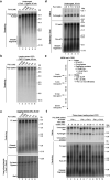
(a) Diagram illustrating the rationale for the oligonucleotide competition assay and the two possible outcomes. CMGE (CMG + Pol ε). (b) Schematic of the possible replication products generated from the oligonucleotide competition assay shown in (c). For simplicity, reciprocal lagging-strand products are not shown. (c) Time course oligonucleotide competition assay in the absence or presence of 5 nM Pol η. (d) Schematic of the possible replication products generated from the oligonucleotide competition assay shown in (e). For simplicity, reciprocal lagging-strand products are not shown. (e) Oligonucleotide competition assay using oligonucleotides mapping to various locations downstream of the CPD (as indicated). Uncropped versions of Fig. 2 gels are available in Source Data Fig. 2.
Fig. 3
Fig. 3. Pol δ, but not Pol ε, inhibits lagging and leading-strand TLS.
(a) Standard replication assay on the lagging-strand CPD template in the presence of 5nM Pol η, Fen1, and Ligase, and increasing concentrations of Pol δ. (b) Standard replication assay on the leading-strand CPD template in the presence of 5 nM Pol η, with increasing concentrations of Pol ε or Pol δ. (c) Mapping of the 3' end of the stalled nascent leading strand in the absence or presence of Pol η and Pol δ. Urea polyacrylamide gel samples were treated with SwaI to generate a stall product and resolved alongside a sequencing ladder. The region surrounding the stalled 3' end of the nascent leading strand is shown to the right of the gel with position of the stall and CPD shown in red. Uncropped versions of Fig. 3 gels are available in Source Data Fig. 3.
Fig. 4
Fig. 4. Pol δ recouples leading-strand synthesis to CMG following TLS.
(a) Standard replication assay on the leading-strand CPD template in the absence or presence of Pol δ, with increasing concentrations of Pol η. (b) Lane profiles of denaturing gel lanes 6 and 12 shown in (a). FL: full-length; UE: uncoupled extension; S: stall; RO: run-off; OF: Okazaki fragments. (c) Reaction scheme for the pulse chase experiment shown in (d). (d) Pulse chase experiment on the leading-strand CPD template in the absence of Pol δ, or with Pol δ (5 nM) added either at the start of the chase or 5 min into the chase. In all cases, 5 nM Pol η was added at the start of the chase. (e-g) Two-dimensional gels of the 20 min time-points in (d). Uncropped versions of Fig. 4 gels are available in Source Data Fig. 4.
Fig. 5
Fig. 5. PCNA monoubiquitination stimulates on the fly TLS
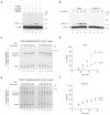
(a) Western blot of PCNA from standard 60 min replication reactions on the leading-strand CPD template. Individual ubiquitination machinery components and MCM10 were omitted from reactions as indicated. (b) Western blot of PCNA from standard time course reactions performed on the leading-strand CPD template with either wild-type PCNA or PCNAK164R in the presence of ubiquitin, Uba1, and Rad6–Rad18. (c) Standard replication reaction time course performed with wild-type PCNA in the absence or presence of Uba1. Reactions contained Pol η, Pol δ, ubiquitin, Rad6–Rad18, Fen1 and Ligase, in addition to standard replication proteins. (d) Quantification of the percentage of bypass in the absence or presence of uba1 as performed in (c). Data are plotted as the means of three independent experiments with error bars representing the SEM. (e) Standard replication reaction time course as performed in (c) but with PCNAK164R. (f) Quantification of the percentage of bypass in the absence or presence of uba1 as performed in (e). Data are plotted as the means of three independent experiments with error bars representing the SEM. Uncropped versions of Fig. 5 gels are available in Source Data Fig. 5.
Fig. 6
Fig. 6. PCNA monoubiquitination promotes lagging-strand TLS.
(a) Standard replication reaction time course performed with wild-type PCNA in the absence or presence of Uba1 on the lagging-strand CPD template. Reactions contained Pol η, Pol δ, ubiquitin, Rad6–Rad18, Fen1 and Ligase, in addition to standard replication proteins. (b) Quantification of the percentage of bypass in the absence or presence of Uba1 as performed in (a). Data are plotted as the means of three independent experiments with error bars representing the SEM. (c) Standard replication reaction time course as performed in (A) but with PCNAK164R. (d) Quantification of the percentage of bypass in the absence or presence of Uba1 as performed in (c). Data are plotted as the means of three independent experiments with error bars representing the SEM. Uncropped versions of Fig. 6 gels are available in Source Data Fig. 6.
Fig. 7
Fig. 7. Model of leading- and lagging-strand TLS by Pol η.

References

    1. Daigaku Y, et al. A global profile of replicative polymerase usage. Nat Struct Mol Biol. 2015;22:192–198. - PMC - PubMed
    1. Nick McElhinny SA, Gordenin DA, Stith CM, Burgers PMJ, Kunkel TA. Division of Labor at the Eukaryotic Replication Fork. Mol Cell. 2008;30:137–144. - PMC - PubMed
    1. Pursell ZF, Isoz I, Lundström E-B, Johansson E, Kunkel TA. Yeast DNA Polymerase ε Participates in Leading-Strand DNA Replication. Science. 2007;317:127–130. - PMC - PubMed
    1. Zeman MK, Cimprich KA. Causes and consequences of replication stress. Nat Cell Biol. 2014;16:2–9. - PMC - PubMed
    1. Marians KJ. Lesion Bypass and the Reactivation of Stalled Replication Forks. Annu Rev Biochem. 2018;87:217–238. - PMC - PubMed

Publication types

MeSH terms