. 2020 Apr 21;14(2):024114.
doi: 10.1063/1.5144617. eCollection 2020 Mar.

Ultrasound-induced molecular delivery to erythrocytes using a microfluidic system

Affiliations

Ultrasound-induced molecular delivery to erythrocytes using a microfluidic system

Connor S Centner et al. Biomicrofluidics. .

Abstract

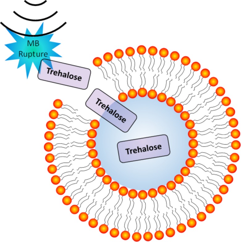
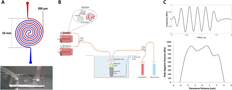
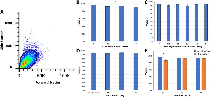
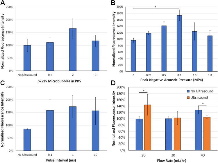
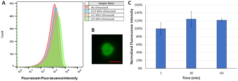
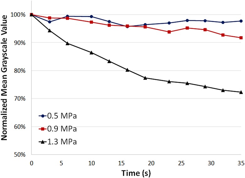
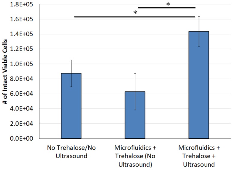
Preservation of erythrocytes in a desiccated state for storage at ambient temperature could simplify blood transfusions in austere environments, such as rural clinics, far-forward military operations, and during space travel. Currently, storage of erythrocytes is limited by a short shelf-life of 42 days at 4 °C, and long-term preservation requires a complex process that involves the addition and removal of glycerol from erythrocytes before and after storage at -80 °C, respectively. Natural compounds, such as trehalose, can protect cells in a desiccated state if they are present at sufficient levels inside the cell, but mammalian cell membranes lack transporters for this compound. To facilitate compound loading across the plasma membrane via ultrasound and microbubbles (sonoporation), a polydimethylsiloxane-based microfluidic device was developed. Delivery of fluorescein into erythrocytes was tested at various conditions to assess the effects of parameters such as ultrasound pressure, ultrasound pulse interval, microbubble dose, and flow rate. Changes in ultrasound pressure and mean flow rate caused statistically significant increases in fluorescein delivery of up to 73 ± 37% (p < 0.05) and 44 ± 33% (p < 0.01), respectively, compared to control groups, but no statistically significant differences were detected with changes in ultrasound pulse intervals. Following freeze-drying and rehydration, recovery of viable erythrocytes increased by up to 128 ± 32% after ultrasound-mediated loading of trehalose compared to control groups (p < 0.05). These results suggest that ultrasound-mediated molecular delivery in microfluidic channels may be a viable approach to process erythrocytes for long-term storage in a desiccated state at ambient temperatures.


Figures

FIG. 1.
FIG. 1.
Illustration of ultrasound-induced microbubble (MB) rupture leading to transient perforation of cell membranes—enabling enhanced intracellular delivery of soluble compounds such as trehalose (not to scale).
FIG. 2.
FIG. 2.
(a) Top: Microfluidic device design with 200 μm channel width. Bottom: a photo of one of the microfluidic devices. (b) Schematic of experimental setup (not to scale). (c) Top: Ultrasound waveform transmitted by the ultrasound probe (ATL P4-1). Bottom: Transverse profile of the ultrasound beam (−3 dB beam width = ∼5 mm).
FIG. 3.
FIG. 3.
(a) Representative flow cytometry scatterplot with the gate used to detect viable erythrocytes based on forward and side scatter profiles. High viability of erythrocytes was observed at (b) different microbubble concentrations, (c) different acoustic pressures, (d) different pulse intervals, and (e) different microfluidic flow rates. Statistically significant decreases in viability were detected at a microbubble dose of 9% (ANOVA p < 0.05) and with ultrasound treatment at a microfluidic flow rate of 20 ml/h (p < 0.05) compared to the no ultrasound control groups.
FIG. 4.
FIG. 4.
(a) Effect of microbubble concentrations on ultrasound-mediated fluorescein uptake. There was a trend toward statistical significance (ANOVA p = 0.12, n = 4/group). (b) Effect of peak negative acoustic pressure on ultrasound-mediated fluorescein uptake. There were statistically significant differences with ultrasound pressures at 0.5 and 0.9 MPa compared to the 0 MPa (no ultrasound) control group (ANOVA p = 0.01, n = 4–7/group). (c) Effect of pulse interval on ultrasound-mediated fluorescein uptake. There were no statistically significant differences between the different pulse intervals (n = 4–5/group). (d) Effect of microfluidic flow rate on ultrasound-mediated fluorescein uptake, ultrasound treatment, and flow rate effect fluorescein uptake, respectively (p < 0.05, n = 5/group).
FIG. 5.
FIG. 5.
(a) Representative fluorescence intensity histograms after ultrasound treatment at peak negative pressures of 0.25 MPa, 0.5 MPa, 0.9 MPa, or no ultrasound (negative control). Fluorescence intensity increased up to peak negative acoustic pressures of 0.9 MPa indicating enhance molecular delivery to erythrocytes. (b) Representative fluorescence microscopy image of an erythrocyte after ultrasound treatment at a peak negative acoustic pressure of 0.9 MPa demonstrating fluorescein delivery (scale bar = 5 μm). (c) Fluorescence intensity of erythrocytes over time after fluorescein treatment as measured by flow cytometry, indicating no significant difference over time (n = 3/group).
FIG. 6.
FIG. 6.
Ultrasound image mean gray scale intensity as a function of time at peak negative acoustic pressures of 0.5 MPa, 0.9 MPa, and 1.3 MPa. At higher pressures (1.3 MPa), the mean gray scale value decreases more rapidly indicating higher rates of microbubble destruction compared to lower pressures (0.5 MPa and 0.9 MPa).
FIG. 7.
FIG. 7.
Number of intact viable erythrocytes following freeze-drying and rehydration. Microfluidic ultrasound treatment with trehalose caused a statistically significant increase in the recovery of intact viable cells compared to the control groups (p < 0.05, n = 4–8/group).

References

    1. Pfuntner A., Wier L. M., and Stocks C., “Most frequent procedures performed in U.S. hospitals, 2011: Statistical brief #165,” in Healthcare Cost and Utilization Project (HCUP) Statistical Briefs (Agency for Healthcare Research and Quality, Rockville, MD, 2006). - PubMed
    1. Haradin A. R., Weed R. I., and Reed C. F., “Changes in physical properties of stored erythrocytes relationship to survival in vivo,” Transfusion 9(5), 229–237 (1969). 10.1111/j.1537-2995.1969.tb04929.x - DOI - PubMed
    1. Izzo P. et al., “Erythrocytes stored in CPD SAG-mannitol: Evaluation of their deformability,” Clin. Hemorheol. Microcirc. 21(3–4), 335–339 (1999). - PubMed
    1. Bunn H. F. et al., “Hemoglobin function in stored blood,” J. Clin. Invest. 48(2), 311–321 (1969). 10.1172/JCI105987 - DOI - PMC - PubMed
    1. Greenwalt T. J., Zehner Sostok C., and Dumaswala U. J., “Studies in red blood cell preservation. 2. Comparison of vesicle formation, morphology, and membrane lipids during storage in AS-1 and CPDA-1,” Vox Sang. 58(2), 90–93 (1990). 10.1111/j.1423-0410.1990.tb02068.x - DOI - PubMed

LinkOut - more resources