

Medial Opening Wedge High Tibial Osteotomy Decreases Medial Meniscal Extrusion and Improves Clinical Outcomes and Return to Activity
- PMID: 32341931
- PMCID: PMC7168781
- DOI: 10.1177/2325967120913531
Medial Opening Wedge High Tibial Osteotomy Decreases Medial Meniscal Extrusion and Improves Clinical Outcomes and Return to Activity
Abstract
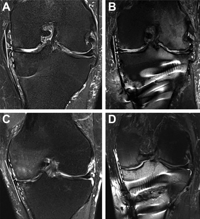
Background: Medial meniscal extrusion (MME) is defined as displacement of the meniscus that extends beyond the tibial margin. Knee varus malalignment increases MME.
Purpose/hypothesis: The purpose of this study was to quantify MME before and after medial opening wedge high tibial osteotomy (HTO) and to correlate the reduction of MME with clinical outcomes and return to activity. It was hypothesized that MME would decrease after HTO and that patients with lower MME after surgery would have improved clinical outcomes and return to activity at short-term follow-up.
Study design: Case series; Level of evidence, 4.
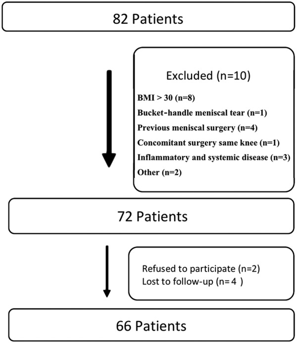
Methods: This study included 66 patients who underwent HTO to correct the anatomic axis with a minimum follow-up of 2 years. MME was measured using magnetic resonance imaging preoperatively and 6 weeks after surgery (study protocol). Patients were assessed preoperatively and postoperatively with the Knee injury and Osteoarthritis Outcome Score (KOOS), visual analog scale (VAS) score for pain, and Tegner score.
Results: The mean ± SD preoperative and postoperative MME values were 3.9 ± 0.6 mm and 0.9 ± 0.5 mm, respectively. At 2 years after surgery, KOOS, pain VAS, and Tegner scores were higher than those found preoperatively (P < .001). Patients with less than 1.5 mm of MME after surgery had better clinical outcomes and return to activity compared with patients who had MME of 1.5 mm or more (P < .05).
Conclusion: Medial opening wedge HTO decreased MME after 6 weeks and improved clinical outcomes and return to activity at a minimum 2-year follow-up. Additionally, patients with postoperative MME of less than 1.5 mm had better clinical outcomes and return to activity compared with patients who had postoperative MME of 1.5 mm or more.
Keywords: high tibial osteotomy; knee function; meniscal extrusion; return to activities.
© The Author(s) 2020.
Conflict of interest statement
The authors declared that there are no conflicts of interest in the authorship and publication of this contribution. AOSSM checks author disclosures against the Open Payments Database (OPD). AOSSM has not conducted an independent investigation on the OPD and disclaims any liability or responsibility relating thereto.
Figures
Comment in
-
High Tibial Osteotomy Decreases Medial Meniscal Extrusion and Improves Clinical Outcomes and Return to Activity: Letter to the Editor.Orthop J Sports Med. 2020 Sep 30;8(9):2325967120953073. doi: 10.1177/2325967120953073. eCollection 2020 Sep. Orthop J Sports Med. 2020. PMID: 33088841 Free PMC article. No abstract available.
References
-
- Achtnich A, Petersen W, Willinger L, et al. Medial meniscus extrusion increases with age and BMI and is depending on different loading conditions. Knee Surg Sports Traumatol Arthrosc. 2018;26(8):2282–2288. - PubMed
-
- Bastard C, Mirouse G, Potage D, et al. Return to sports and quality of life after high tibial osteotomy in patients under 60 years of age. Orthop Traumatol Surg Res. 2017;103(8):1189–1191. - PubMed
-
- Boxheimer L, Lutz AM, Treiber K, Goepfert K, Crook DW. MR imaging of the knee: position related changes of the menisci in asymptomatic volunteers. Invest Radiol. 2004;39:254–263. - PubMed
LinkOut - more resources
Full Text Sources
Miscellaneous
