. 2020 Apr 22;6(4):546-554.
doi: 10.1021/acscentsci.9b01268. Epub 2020 Apr 13.

Light-Activatable, 2,5-Disubstituted Tetrazoles for the Proteome-wide Profiling of Aspartates and Glutamates in Living Bacteria

Affiliations

Light-Activatable, 2,5-Disubstituted Tetrazoles for the Proteome-wide Profiling of Aspartates and Glutamates in Living Bacteria

Kathrin Bach et al. ACS Cent Sci. .

Abstract

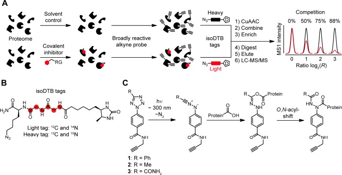
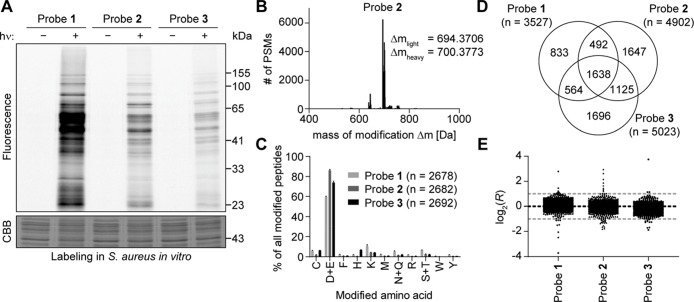
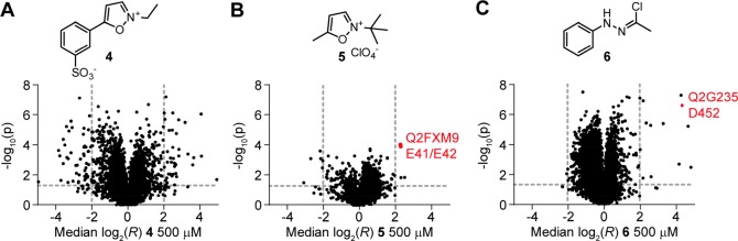
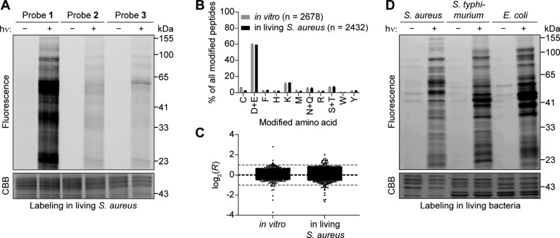
Covalent inhibitors have recently seen a resurgence of interest in drug development. Nevertheless, compounds, which do not rely on an enzymatic activity, have almost exclusively been developed to target cysteines. Expanding the scope to other amino acids would be largely facilitated by the ability to globally monitor their engagement by covalent inhibitors. Here, we present the use of light-activatable 2,5-disubstituted tetrazoles that allow quantifying 8971 aspartates and glutamates in the bacterial proteome with excellent selectivity. Using these probes, we competitively map the binding sites of two isoxazolium salts and introduce hydrazonyl chlorides as a new class of carboxylic-acid-directed covalent protein ligands. As the probes are unreactive prior to activation, they allow global profiling even in living Gram-positive and Gram-negative bacteria. Taken together, this method to monitor aspartates and glutamates proteome-wide will lay the foundation to efficiently develop covalent inhibitors targeting these amino acids.


Conflict of interest statement

The authors declare no competing financial interest.

Figures

Figure 1
Figure 1
Concept of this study. (A) Workflow of competitive residue-specific proteomics using the isotopically labeled desthiobiotin azide (isoDTB) tags. RG, reactive group; D, desthiobiotin. (B) Structure of the isoDTB tags. (C) Light-induced reactivity of 2,5-disubstituted tetrazoles 13 with carboxylic acids in proteins. While other nucleophiles might attack the nitrilimine, only for carboxylic acids a stable product can be formed via an O,N-acyl-shift.
Figure 2
Figure 2
Light-activatable 2,5-disubstituted tetrazoles allow global monitoring of aspartates and glutamates in the S. aureus proteome in vitro with high specificity. (A) Gel-based analysis of labeling with probes 13. S. aureus lysate was treated with 100 μM of the indicated probe, incubated for 30 min, irradiated with light (λ = 280–315 nm) for 10 min, and labeled with TAMRA-azide using CuAAC. Controls were performed without irradiation. Gel-based analysis was performed with in-gel fluorescence scanning and staining using Coomassie Brilliant Blue (CBB). (B) Analysis of the mass of modification on tryptic peptides after labeling of S. aureus lysate with 100 μM probe 2. MSFragger software was used to determine, which masses of modification occur in the proteomic samples labeled with probe 2 after light activation and CuAAC to the light and heavy isoDTB tags. Expected masses of modification for tryptic peptides labeled with 2 according to the reactivity shown in Figure 1C and additionally modified with light or heavy isoDTB tag, respectively, are 694.3663 and 700.3738 Da. PSM: peptide-spectrum match. (C) Analysis of the amino acid specificity of the probes. Proteomes labeled with the indicated probe after light activation and modified by CuAAC with the light and heavy isoDTB tags were analyzed with MaxQuant software allowing the modification on any potentially nucleophilic amino acid. Peptides were included in the analysis if the localization probability for a single residue was more than 75%. Data shows the mean ± the standard deviation. The total number of identified PSMs is given in parentheses. (D) Venn diagram of the number of quantified aspartates and glutamates with the three different probes. (E) Plot of the ratios log2(R) for aspartates and glutamates in proteomic samples, in which the heavy- and light-labeled sample were both modified with 100 μM of the indicated probe without pretreatment with an inhibitor. The expected value of log2(R) = 0 is indicated by the black line; the preferred quantification window (−1 < log2(R) < 1) is indicated by the two gray lines. Each dot represents one quantified aspartate or glutamate. All data for panels B–E originates from biologically independent duplicates of technical duplicates.
Figure 3
Figure 3
Probe 2 reveals the targeted residues of carboxylic-acid-directed covalent protein ligands proteome-wide using isoDTB-ABPP in vitro. (A–C) Volcano plots of isoDTB-ABPP experiments comparing samples pretreated with 500 μM of the indicated covalent ligand to a solvent control. Plotted are the ratio (log2(R)) between the heavy (solvent-treated, 1% HCl for 4, DMSO for 5, DMF for 6) and light (compound-treated) labeled samples and the probability in a one-sample t test that R is equal to one (−log10(p)). The targeted E41/E42 of pyruvate kinase (UniProt code: Q2FXM9) and D452 of nicotinate phosphoribosyltransferase (UniProt code: Q2G235) are highlighted in red. All data originates from two (B and C) or three (A) biologically independent experiments; performed in technical duplicates (A and B) or triplicates (C).
Figure 4
Figure 4
Specific labeling of aspartates and glutamates with 2,5-disubstituted tetrazoles in living bacteria. (A) Probe 1 efficiently labels proteins in living S. aureus. Living bacteria were treated with the indicated probe for 1 h and labeled by irradiation with light (λ = 280–315 nm) for 10 min. After lysis, TAMRA azide was attached using CuAAC, and labeling was analyzed using gel electrophoresis with in-gel fluorescence scanning and Coomassie Brilliant Blue (CBB) staining. Controls were performed without irradiation. (B) Analysis of the amino acid specificity of probe 1in vitro and in living S. aureus. Samples were analyzed with MaxQuant software allowing the modification with probe 1 and the heavy or light isoDTB tag to be on any potentially nucleophilic amino acid. Peptides were further analyzed if the localization probability for a single residue was more than 75%. The total number of identified modification sites is given in parentheses. Data shows the mean ± the standard deviation. (C) Plot of the ratios log2(R) of samples, in which the heavy- and light-labeled sample were both modified with the same concentration of the indicated probe in living S. aureus without pretreatment with an inhibitor. The expected value of log2(R) = 0 is indicated by the black line; the preferred quantification window (−1 < log2(R) < 1) is indicated by the two gray lines. One data point in living bacteria at log2(R) = −8.9 is not shown for clarity. All data for panels B and C originates from biologically independent duplicates of technical triplicates for data in living bacteria and from biologically independent duplicates of technical duplicates for data in vitro. (D) Probe 1 efficiently labels proteins in the living Gram-negative bacteria S. typhimurium and E. coli. The experiment was performed in the indicated bacteria as described for S. aureus in part A.

Similar articles

Cited by

References

    1. Singh J.; Petter R. C.; Baillie T. A.; Whitty A. The resurgence of covalent drugs. Nat. Rev. Drug Discovery 2011, 10, 307–317. 10.1038/nrd3410. - DOI - PubMed
    1. Chaikuad A.; Koch P.; Laufer S. A.; Knapp S. The Cysteinome of Protein Kinases as a Target in Drug Development. Angew. Chem., Int. Ed. 2018, 57, 4372–4385. 10.1002/anie.201707875. - DOI - PubMed
    1. Hendlin D.; Stapley E. O.; Jackson M.; Wallick H.; Miller A. K.; Wolf F. J.; Miller T. W.; Chaiet L.; Kahan F. M.; Foltz E. L.; Woodruff H. B.; Mata J. M.; Hernandez S.; Mochales S. Phosphonomycin, a new antibiotic produced by strains of streptomyces. Science 1969, 166, 122–123. 10.1126/science.166.3901.122. - DOI - PubMed
    1. Nishimura H.; Mayama M.; Komatsu Y.; Kato H.; Shimaoka N.; Tanaka Y. Showdomycin, a New Antibiotic from a Streptomyces Sp. J. Antibiot. 1964, 17, 148–155. - PubMed
    1. Smith P. A.; Koehler M. F. T.; Girgis H. S.; Yan D.; Chen Y.; Chen Y.; Crawford J. J.; Durk M. R.; Higuchi R. I.; Kang J.; Murray J.; Paraselli P.; Park S.; Phung W.; Quinn J. G.; Roberts T. C.; Rouge L.; Schwarz J. B.; Skippington E.; Wai J.; Xu M.; Yu Z.; Zhang H.; Tan M. W.; Heise C. E. Optimized arylomycins are a new class of Gram-negative antibiotics. Nature 2018, 561, 189–194. 10.1038/s41586-018-0483-6. - DOI - PubMed