Observational Study
. 2021 Feb;73(2):759-775.
doi: 10.1002/hep.31297. Epub 2020 Nov 2.

Growth Hormone Stops Excessive Inflammation After Partial Hepatectomy, Allowing Liver Regeneration and Survival Through Induction of H2-Bl/HLA-G

Affiliations
Observational Study

Growth Hormone Stops Excessive Inflammation After Partial Hepatectomy, Allowing Liver Regeneration and Survival Through Induction of H2-Bl/HLA-G

Mayumi Ishikawa et al. Hepatology. 2021 Feb.

Abstract

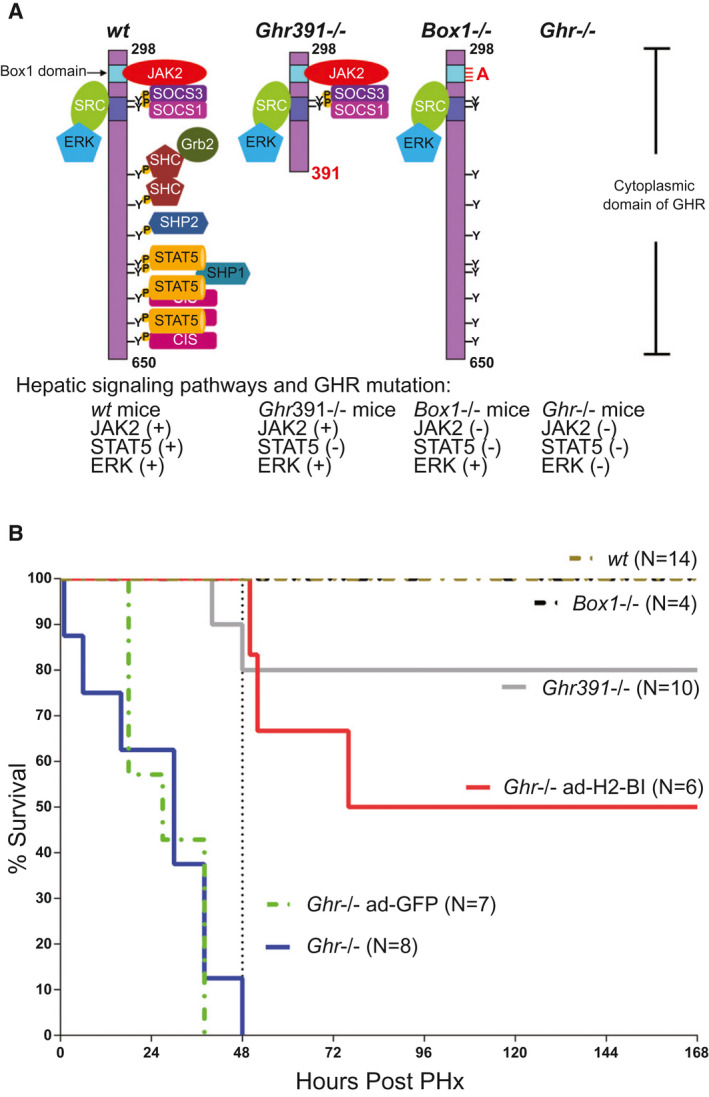
Background and aims: Growth hormone (GH) is important for liver regeneration after partial hepatectomy (PHx). We investigated this process in C57BL/6 mice that express different forms of the GH receptor (GHR) with deletions in key signaling domains.

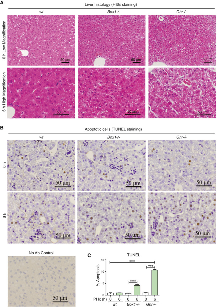
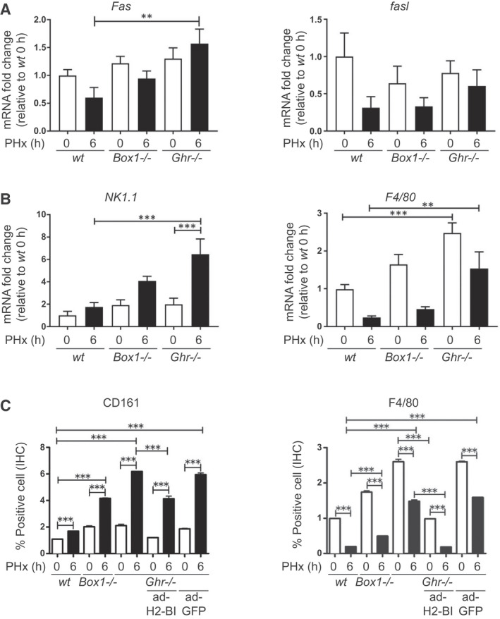
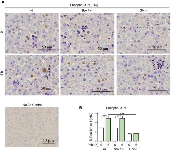
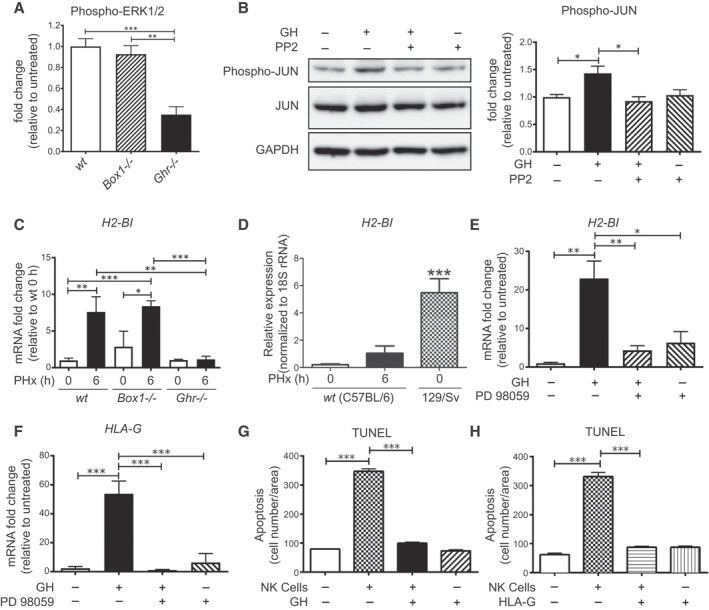
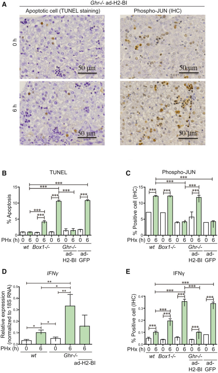
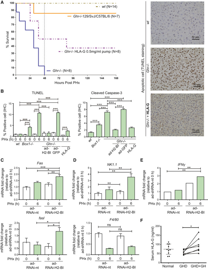
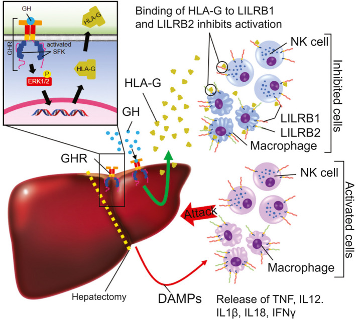
Approach and results: PHx was performed on C57BL/6 mice lacking GHR (Ghr-/- ), disabled for all GH-dependent Janus kinase 2 signaling (Box1-/- ), or lacking only GH-dependent signal transducer and activator of transcription 5 (STAT5) signaling (Ghr391-/- ), and wild-type littermates. C57BL/6 Ghr-/- mice showed striking mortality within 48 hours after PHx, whereas Box1-/- or Ghr391-/- mice survived with normal liver regeneration. Ghr-/- mortality was associated with increased apoptosis and elevated natural killer/natural killer T cell and macrophage cell markers. We identified H2-Bl, a key immunotolerance protein, which is up-regulated by PHx through a GH-mediated, Janus kinase 2-independent, SRC family kinase-dependent pathway. GH treatment was confirmed to up-regulate expression of the human homolog of H2-Bl (human leukocyte antigen G [HLA-G]) in primary human hepatocytes and in the serum of GH-deficient patients. We find that injury-associated innate immune attack by natural killer/natural killer T cell and macrophage cells are instrumental in the failure of liver regeneration, and this can be overcome in Ghr-/- mice by adenoviral delivery of H2-Bl or by infusion of HLA-G protein. Further, H2-Bl knockdown in wild-type C57BL/6 mice showed elevated markers of inflammation after PHx, whereas Ghr-/- backcrossed on a strain with high endogenous H2-Bl expression showed a high rate of survival following PHx.

Conclusions: GH induction of H2-Bl expression is crucial for reducing innate immune-mediated apoptosis and promoting survival after PHx in C57BL/6 mice. Treatment with HLA-G may lead to improved clinical outcomes following liver surgery or transplantation.


Figures

FIG. 1
FIG. 1
Survival of Ghr mutant C57BL/6 mouse strains after PHx. (A) Signaling capabilities of GHR mutant mice. The cytoplasmic domain of receptor is shown, with remaining signaling pathways for each mutant. Ghr391 −/− eliminates STAT5 signaling; Box1 −/− removes all JAK/STAT signaling while maintaining SFK signaling; and Ghr −/− removes all GH mediated signaling. (B) Survival of mouse strains after PHx (number of mice for each genotype shown), in which the mortality of Ghr −/− is partially rescued by adenoviral expression of H2Bl. Abbreviations: CIS, cytokine‐inducible SH2 (Src homology 2) protein; SHC, Src Homology and Collagen protein; SHP1, Src homology region 2 domain‐containing phosphatase‐1; SHP2, Src homology region 2 domain‐containing phosphatase‐2.
FIG. 2
FIG. 2
The livers of Ghr −/− mice 6 hours following PHx show lipid droplets, ballooning, and increased apoptosis compared with wt and Box −/− mice. (A) Liver histology from Box1 −/− and Ghr −/− mice compared with wt 6 hours after PHx, showing lipid droplets and ballooning in Ghr −/− liver. Lower panel shows high magnification images. (B) TUNEL staining in livers at 0 hours and 6 hours after PHx, showing increased apoptosis in Box1 −/− mice compared with wt, but particularly in Ghr −/− mice compared with wt. Staining without primary antibody is shown (No Ab Control). (C) TUNEL quantification in livers at 0 hours and 6 hours after PHx. Six to nine sections were analyzed for each mouse at 0 hours and 6 hours following PHx, with n = 4‐11 mice per group. Abbreviation: H&E, hematoxylin and eosin.
FIG. 3
FIG. 3
Immune attack is elevated in Ghr −/− mice compared with wt mice following PHx, and this elevation can be ameliorated by expression of H2Bl. (A) Fas and fasl transcripts at 0 hours and 6 hours after PHx, showing significantly elevated Fas transcript relative to wt in Ghr −/− mice at 6 hours. (B) NK/NKT marker NK1.1 and macrophage marker F4/80 transcripts in livers at 0 hours and 6 hours after PHx, showing increased level of NK/NKT marker 6 hours after PHx in Ghr −/− mice, and elevated macrophage marker at both 0 hours and 6 hours in Ghr −/− relative to wt. (C) Protein expression of NK1.1 and F4/80 by IHC, showing correspondence with transcript levels. Also shown is the ability of ad‐H2‐Bl, but not ad‐GFP, to reverse these changes in Ghr −/− mice (n = 4‐6 mice per group).
FIG. 4
FIG. 4
JUN is activated in livers of wt and Box1 −/− mice after PHx, but not in Ghr −/− mice. (A) JUN activation/phosphorylation is deficient in Ghr −/− mice, but not in wt or Box1 −/− mice, 6 hours after PHx. Representative high‐magnification IHC sections are shown. Staining without primary antibody is shown (No Ab Control). (B) Quantification of large nuclei with phospho‐JUN immunoreactivity is shown (five sections per mouse, n = 3 mice per group).
FIG. 5
FIG. 5
GH activates JUN and up‐regulates H2Bl and HLA‐G expression in a SRC‐ERK pathway–dependent manner, protects hepatocytes from NK cell attack, and H2‐Bl expression is induced in livers of wt and Box1 −/− C57BL/6 mice after PHx, but not in Ghr −/− C57BL/6 mice. (A) ERK1/2 activation in Ghr −/− liver 6 hours after PHx is impaired compared with Box1 −/− and wt mice (quantification from western blot, n = 3 for each mouse line). (B) Immunoblot showing suppression of GH‐stimulated JUN phosphorylation by SRC kinase inhibitor PP2 (10 μM) in AML12 hepatocyte cells and its quantification (n = 3 wells per condition). (C) H2‐Bl transcript level does not increase after PHx in Ghr −/− , whereas wt and Box1 −/− show a significant increase (n = 3‐6 per group). (D) Basal expression of H2Bl transcript in the 129/Sv mouse strain is high compared with C57BL/6. (E) GH treatment increases H2‐Bl transcript in AML12 mouse hepatocyte cells, and this increase is blocked by the MEK inhibitor PD98059 (20 μM) (n = 3 replicates per group). Inhibition of ERK1/2 is shown in Supporting Fig. S3. (F) GH treatment increases HLA‐G transcript in human hepatocyte cells, and this increase is blocked by the MEK inhibitor PD98059 (20 μM) (n = 3 replicates per group). Quantification of TUNEL staining of human hepatocytes following co‐culture with human NK cells and treatment with either GH (G) or HLA‐G (H) protein. Abbreviations: GAPDH, glyceraldehyde 3‐phosphate dehydrogenase; PP2, pyrazolopyrimidine (PP)2; and rRNA, ribosomal RNA.
FIG. 6
FIG. 6
Indicators of immune attack are reversed by adenoviral delivery of H2Bl in Ghr −/− mice. (A) TUNEL and phospho‐JUN staining shows ability of ad‐H2‐Bl to prevent apoptosis and restore phospho‐JUN level in Ghr −/− mice at 6 hours following PHx. Quantification of TUNEL (B) and phospho‐JUN staining (C), five sections per mouse (n = 3 mice/group). (D) The increase in IFNγ transcript levels after PHx is higher in Ghr −/− mice compared with wt mice, and this increase in IFNγ transcript in Ghr −/− is reduced by adenoviral expression of H2Bl. (E) IFNγ expression by IHC shows increased expression at 6 hours in wt, but 3.5‐fold higher expression in Ghr −/− , which is markedly decreased by ad‐H2‐Bl but not by ad‐GFP (five sections per mouse, n = 3 mice/group).
FIG. 7
FIG. 7
Treatment with HLA‐G or mixed background mice with high expression of H2Bl improves survival of Ghr −/− mice, while markers of immune attack are elevated in C57BL/6 mice by knockdown of H2‐Bl following PHx. (A) Survival of mouse strains after PHx (number for each genotype shown) and osmotic pump infusion of HLA‐G. (B) Infusion of HLA‐G protein markedly reduced apoptosis levels in Ghr −/− mice. TUNEL staining showing ability of HLA‐G to prevent apoptosis in Ghr −/− mice at 6 hours PHx with representative images shown (right). Quantification of TUNEL and cleaved caspase‐3, 5 sections per mouse (n = 3 mice per group). (C) Transcripts analysis in liver at 6 hours after PHx with H2‐Bl RNAi show elevated levels of Fas and fasl compared to non‐targeting control. (D) NK/NKT marker NK1.1 is elevated at 6 hours following PHx with H2‐Bl RNAi compared to non‐targeting RNAi while macrophage marker F4/80 transcripts showed no significant change. (E) IFNγ transcript levels in the liver are increased with H2‐Bl RNAi 6 hours after PHx compared with nontargeting RNAi (n = 6 ad‐RNAi‐H2‐Bl and n = 4 ad‐RNAi‐nt). (F) HLA‐G levels in serum from patients with GHD, patients with GDH following GH treatment, and healthy controls. Abbreviation: ns, not significant.
FIG. 8
FIG. 8
Growth hormone protects the liver from excessive inflammation through SFK‐dependent induction of HLA‐G (H2‐B1 in mice) after PHx. Following PHx surgery, immune cells such as NK cells and macrophages are activated and release inflammatory cytokines, which may contribute to liver failure and mortality. GH induces the expression of HLA‐G, which inhibits immune cell activation, keeping inflammation in check, which permits regeneration.

Comment in

References

    1. Gao B, Radaeva S, Park O. Liver natural killer and natural killer T cells: immunobiology and emerging roles in liver diseases. J Leukoc Biol 2009;86:513‐528. - PMC - PubMed
    1. Notas G, Kisseleva T, Brenner D. NK and NKT cells in liver injury and fibrosis. Clin Immunol 2009;130:16‐26. - PubMed
    1. Pennisi PA, Kopchick JJ, Thorgeirsson S, LeRoith D, Yakar S. Role of growth hormone (GH) in liver regeneration. Endocrinology 2004;145:4748‐4755. - PubMed
    1. Yamada Y, Kirillova I, Peschon JJ, Fausto N. Initiation of liver growth by tumor necrosis factor: deficient liver regeneration in mice lacking type I tumor necrosis factor receptor. Proc Natl Acad Sci U S A 1997;94:1441‐1446. - PMC - PubMed
    1. Cressman DE, Greenbaum LE, DeAngelis RA, Ciliberto G, Furth EE, Poli V, et al. Liver failure and defective hepatocyte regeneration in interleukin‐6‐deficient mice. Science 1996;274:1379‐1383. - PubMed

Publication types

MeSH terms