. 2020 May 29;40(5):BSR20192549.
doi: 10.1042/BSR20192549.

ADSC-Exos containing MALAT1 promotes wound healing by targeting miR-124 through activating Wnt/β-catenin pathway

Affiliations

ADSC-Exos containing MALAT1 promotes wound healing by targeting miR-124 through activating Wnt/β-catenin pathway

Lin He et al. Biosci Rep. .

Abstract

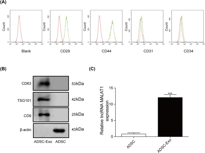
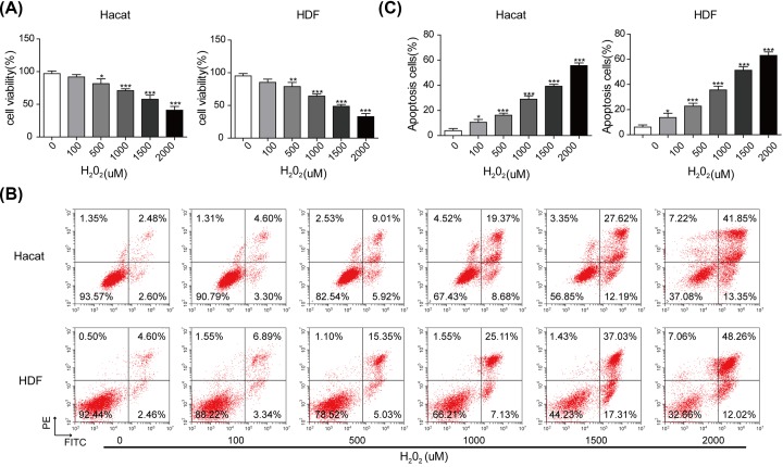
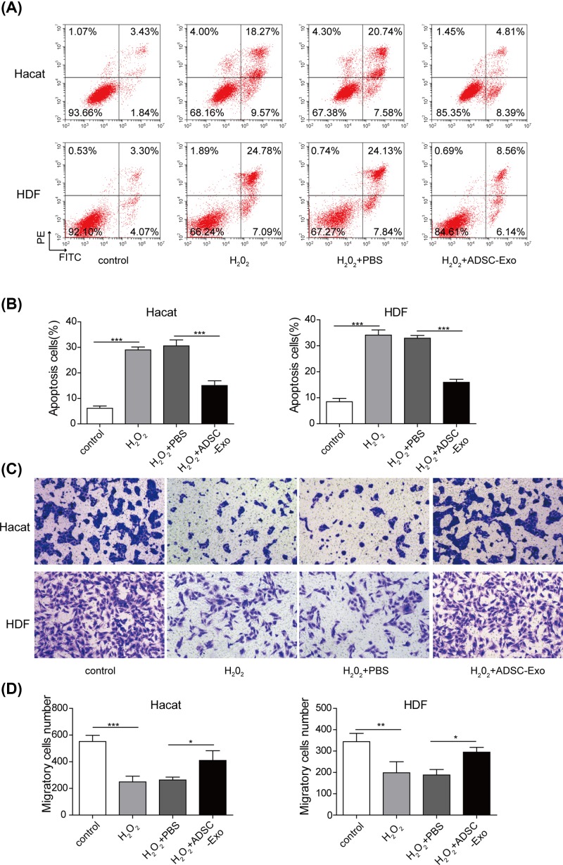
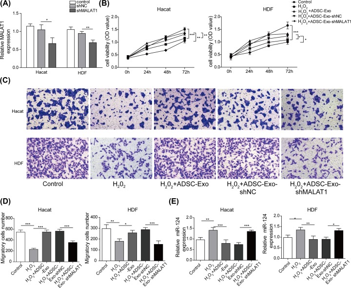
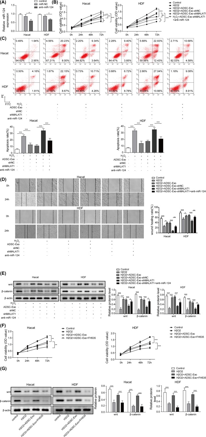
Cutaneous wound is a soft tissue injury that is difficult to heal during aging. It has been demonstrated that adipose-derived stem cells (ADSCs) and its secreted exosomes exert crucial functions in cutaneous wound healing. The present study aimed to elucidate the mechanism of exosomes derived from ADSCs (ADSC-Exos) containing MALAT1 in wound healing. ADSCs were isolated from human normal subcutaneous adipose tissues and identified by flow cytometry analysis. Exosomes were extracted from ADSC supernatants and MALAT1 expression was determined using qRT-PCR analysis. HaCaT and HDF cells were exposed to hydrogen peroxide (H2O2) for simulating the skin lesion model. Subsequently, CCK-8, flow cytometry, wound healing and transwell assays were employed to validate the role of ADSC-Exos containing MALAT1 in the skin lesion model. Besides, cells were transfected with sh-MALAT1 to verify the protective role of MALAT1 in wound healing. The binding relationship between MALAT1 and miR-124 were measured by dual-luciferase reporter assay. ADSC-Exos promoted cell proliferation, migration, and inhibited cell apoptosis of HaCaT and HDF cells impaired by H2O2. However, the depletion of MALAT1 in ADSC-Exos lose these protective effects on HaCaT and HDF cells. Moreover, miR-124 was identified to be a target of MALAT1. Furthermore, ADSC-Exos containing MALAT1 could mediate H2O2-induced wound healing by targeting miR-124 and activating Wnt/β-catenin pathway. ADSC-Exos containing MALAT1 play a positive role in cutaneous wound healing possibly via targeting miR-124 through activating the Wnt/β-catenin pathway, which may provide novel insights into the therapeutic target for cutaneous wound healing.

Keywords: MALAT1; adipose-derived stem cell; cutaneous wound healing; exosomes; miR-124.


Conflict of interest statement

The authors declare that there are no competing interests associated with the manuscript.

Figures

Figure 1
Figure 1. Isolation and characterization of ADSCs and ADSC-Exos
(A) The expression of biomarkers (CD34, CD31, CD29 and CD44) were detected by FACS. (B) The exosomes’ marker proteins (CD63, TSG101 and CD9) detected by Western blot. (C) The expression level of MALAT1 was measured using qRT-PCR assay. ***P<0.001.
Figure 2
Figure 2. Construction of the skin lesion model in vitro
(A) Cell viability assessed by MTT assay. (B) Apoptotic rate of HaCaT and HDF cells exposed with different concentrations of H2O2 examined by flow cytometry assay. (C) The percentage of apoptosis cell for HaCaT and HDF cells. *P<0.05, **P<0.01, ***P<0.001.
Figure 3
Figure 3. Effects of ADSC-Exos on cell proliferation, apoptosis and migration of HaCaT cells impaired by H2O2
(A) Apoptosis of HaCaT and HDF cells was evaluated by flow cytometry assay. (B) The percentage of apoptosis cell for HaCaT and HDF cells. (C,D) The capacity of cell migration in HaCaT and HDF cells was analyzed by transwell assay. *P<0.05, **P<0.01, ***P<0.001.
Figure 4
Figure 4. ADSC-Exos knocking down MALAT1 lose protective effects on HaCaT and HDF cells
(A) The expression level of MALAT1 was measured by qRT-PCR assay. (B) Proliferation of HaCaT and HDF cells treated with H2O2, H2O2+ADSC-Exo, H2O2+ADSC-Exo-shNC or H2O2+ADSC-Exo-shMALAT1 were evaluated by CCK-8 assay. (C,D) Migration of HaCaT and HDF cells treated with H2O2, H2O2+ADSC-Exo, H2O2+ADSC-Exo-shNC or H2O2+ADSC-Exo-shMALAT1 were analyzed by Transwell assay. (E) The expression level of miR-124 was performed using qRT-PCR analysis. *P<0.05, **P<0.01, ***P<0.001.
Figure 5
Figure 5. MALAT1 acts as a sponge of miR-124
(A) Analysis of binding sites of MALAT1 and miR-124 using bioinformatics software. (B) The wild-type and mutant-type of the complementary binding sequences between MALAT1 and miR-124. (C) The binding relationship between MALAT1 and miR-124 was employed using dual-luciferase reporter assay. (D) qRT-PCR was used to detect the expression of miR-124 after transfection with sh-MALAT1. **P<0.01.
Figure 6
Figure 6. ADSC-Exos containing MALAT1 mediates H2O2 induced wound healing by targeting miR-124 through activating Wnt/β-catenin pathway
(A) The expression level of miR-124 was detected by qRT-PCR after transfected with anti-miR-124. (B) CCK-8 assay was performed to assess the cell proliferation of HaCaT and HDF cells treated with H2O2, H2O2+ADSC-Exo, H2O2+ADSC-Exo-shNC, H2O2+ADSC-Exo-shMALAT1, H2O2+ADSC-Exo-shMALAT1+anti-miR-124. (C) Flow cytometry was subjected to evaluate cell apoptosis of HaCaT and HDF cells treated with H2O2, H2O2+ADSC-Exo, H2O2+ADSC-Exo-shNC, H2O2+ADSC-Exo-shMALAT1, H2O2+ADSC-Exo-shMALAT1+anti-miR-124. (D) Migration of HaCaT and HDF cells treated with H2O2, H2O2+ADSC-Exo, H2O2+ADSC-Exo-shNC, H2O2+ADSC-Exo-shMALAT1, H2O2+ADSC-Exo-shMALAT1+anti-miR-124 were analyzed by the scratch wound healing assay. (E) The expression of Wnt/β-catenin signals were determined by western blot. (F) Cell proliferation of HaCaT and HDF cells treated with FH535 were evaluated by CCK-8 assay. (G) Wnt/β-catenin signal pathway of HaCaT and HDF cells treated with H2O2, H2O2+ADSC-Exo or H2O2+ADSC-Exo+FH535 were monitored by western blot. *P<0.05, **P<0.01, ***P<0.001.

References

    1. Rodriguez-Menocal L., Salgado M., Ford D. and Van Badiavas E. (2012) Stimulation of skin and wound fibroblast migration by mesenchymal stem cells derived from normal donors and chronic wound patients. Stem Cells Transl. Med. 1, 221–229 10.5966/sctm.2011-0029 - DOI - PMC - PubMed
    1. Guo S. and Dipietro L.A. (2010) Factors affecting wound healing. J. Dent. Res. 89, 219–229 10.1177/0022034509359125 - DOI - PMC - PubMed
    1. Zhao P., Sui B.D., Liu N., Lv Y.J., Zheng C.X., Lu Y.B. et al. . (2017) Anti-aging pharmacology in cutaneous wound healing: effects of metformin, resveratrol, and rapamycin by local application. Aging Cell 16, 1083–1093 10.1111/acel.12635 - DOI - PMC - PubMed
    1. Sgonc R. and Gruber J. (2013) Age-related aspects of cutaneous wound healing: a mini-review. Gerontology 59, 159–164 10.1159/000342344 - DOI - PubMed
    1. Lewis C.J., Mardaryev A.N., Poterlowicz K., Sharova T.Y., Aziz A., Sharpe D.T. et al. . (2014) Bone morphogenetic protein signaling suppresses wound-induced skin repair by inhibiting keratinocyte proliferation and migration. J. Invest. Dermatol. 134, 827–837 10.1038/jid.2013.419 - DOI - PMC - PubMed

Publication types

MeSH terms