. 2020 Jun 23;14(6):6519-6531.
doi: 10.1021/acsnano.9b08216. Epub 2020 May 1.

Affinity-Driven Design of Cargo-Switching Nanoparticles to Leverage a Cholesterol-Rich Microenvironment for Atherosclerosis Therapy

Affinity-Driven Design of Cargo-Switching Nanoparticles to Leverage a Cholesterol-Rich Microenvironment for Atherosclerosis Therapy

Heegon Kim et al. ACS Nano. .

Abstract

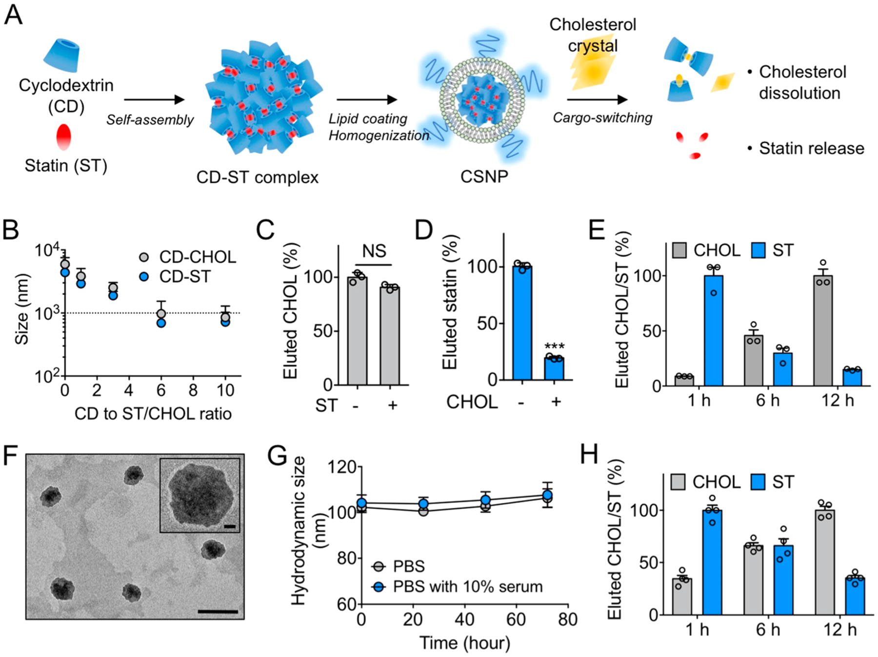
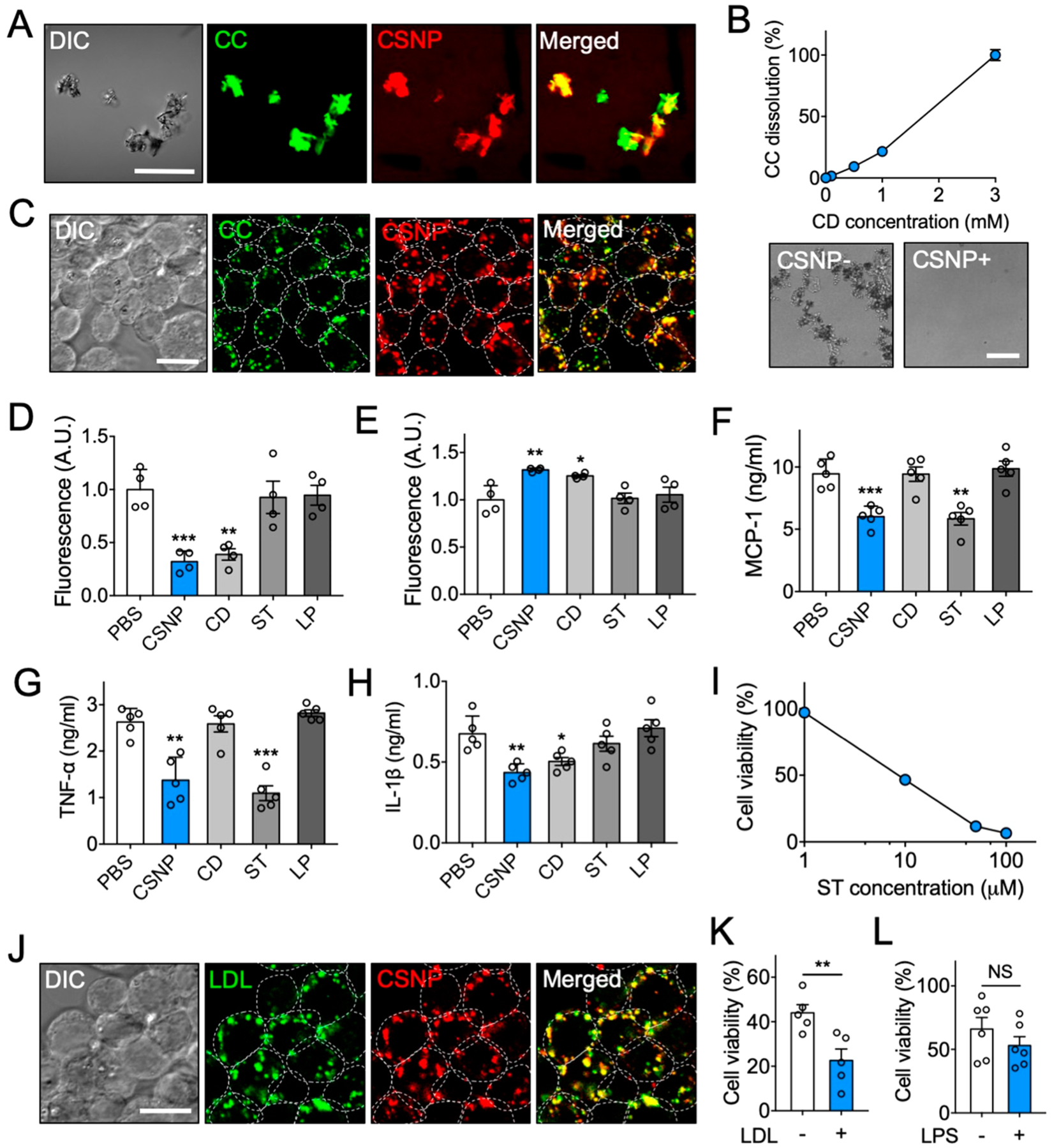
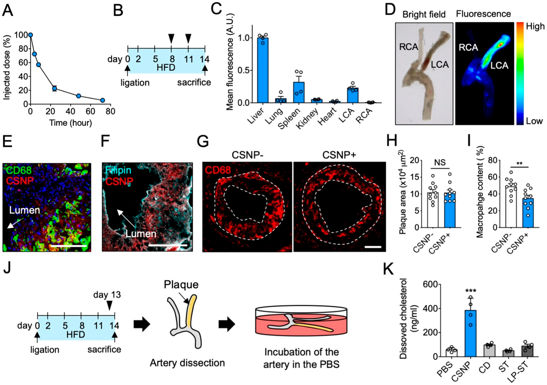
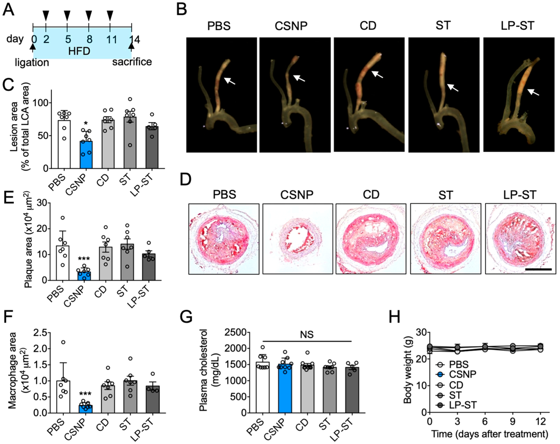
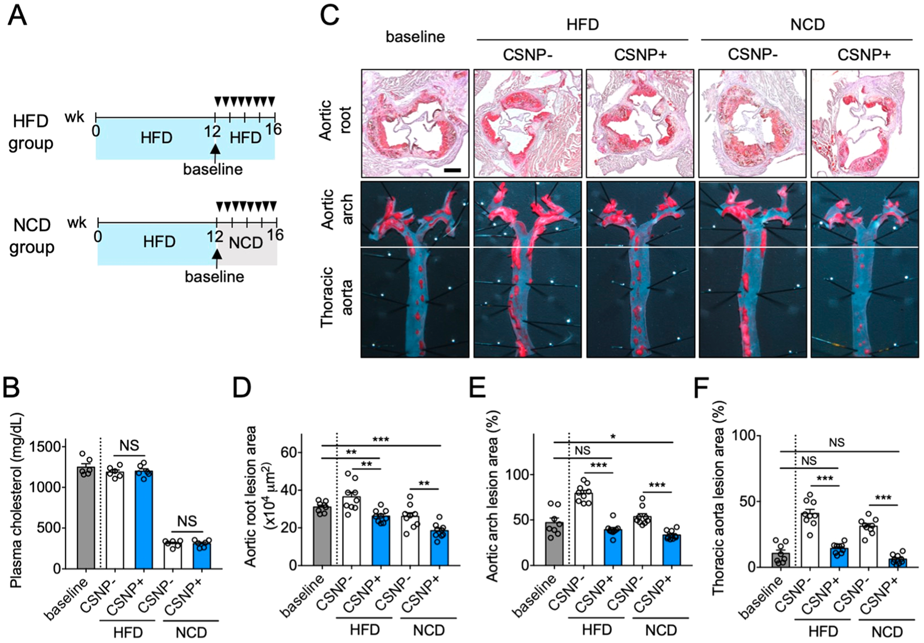
Atherosclerotic plaques exhibit high deposition of cholesterol and macrophages. These are not only the main components of the plaques but also key inflammation-triggering sources. However, no existing therapeutics can achieve effective removal of both components within the plaques. Here, we report cargo-switching nanoparticles (CSNP) that are physicochemically designed to bind to cholesterol and release anti-inflammatory drug in the plaque microenvironment. CSNP have a core-shell structure with a core composed of an inclusion complex of methyl-β-cyclodextrin (cyclodextrin) and simvastatin (statin), and a shell of phospholipids. Upon interaction with cholesterol, which has higher affinity to cyclodextrin than statin, CSNP release statin and scavenge cholesterol instead through cargo-switching. CSNP exhibit cholesterol-sensitive multifaceted antiatherogenic functions attributed to statin release and cholesterol depletion in vitro. In mouse models of atherosclerosis, systemically injected CSNP target atherosclerotic plaques and reduce plaque content of cholesterol and macrophages, which synergistically leads to effective prevention of atherogenesis and regression of established plaques. These findings suggest that CSNP provide a therapeutic platform for interfacing with cholesterol-associated inflammatory diseases such as atherosclerosis.

Keywords: atherosclerosis; cargo-switching; cholesterol; microenvironment; nanoparticle.


Conflict of interest statement

The authors declare no competing financial interest.

Figures

Figure 1.
Figure 1.
Preparation and physicochemical properties of cargo-switching nanoparticles (CSNP). (A) Schematic of CSNP preparation and cargo-switching. (B) Hydrodynamic sizes of cyclodextrin–statin (CD–ST) and cyclodextrin–cholesterol (CD–CHOL) complexes prepared at different CD to ST/CHOL ratios. (C, D) Competitive binding of (C) CHOL and (D) ST to CD. (E) Release of statin and dissolution of cholesterol by CD–ST complexes via cargo-switching. (F) Representative transmission electron microscopic images of CSNP. The scale bar indicates 200 nm. The inset shows a polymer coating layer on the surface of CSNP. The scale bar in the inset indicates 20 nm. (G) Serum stability of CSNP. Hydrodynamic size of CSNP incubated in PBS and PBS containing 10% serum at 37 °C was monitored over a 72-h period. (H) Cargo-switching property of CSNP. The results are representative of three independent experiments. The results are representative of three independent experiments. Data are means ± s.e.m. [n = 5 for B; n = 3 for C and D; n = 4 for G and H; NS, not significant, ***P < 0.001, unpaired two-tailed Student’s t test for C and D].
Figure 2.
Figure 2.
Multiple anti-inflammatory properties of CSNP in vitro. (A) Representative confocal microscopic images of CSNP binding to cholesterol crystals (CC). The scale bar indicates 50 μm. (B) Dose-dependent CC dissolution with CSNP. Bright-field images show complete dissolution of CC 24 h after CSNP treatment at 3 mM CD. (C) Representative confocal microscopic images of CC-laden macrophages after CSNP treatment. The scale bar indicates 10 μm. (D, E) Assessment of cholesterol (D) within cells and (E) in the supernatant after CSNP treatment. (F, G) Secretion of (F) MCP-1 and (G) TNF-α from the LPS-activated macrophages after CSNP treatment. (H) Secretion of IL-1β from the CC-laden LPS-activated macrophages after CSNP treatment. (I) Dose-dependent effects of CSNP on macrophage viability. (J) Representative confocal microscopic images of LDL-laden macrophage after CSNP treatment. The scale bar indicates 10 μm. (K) LDL-dependent effects of CSNP on macrophage viability. (L) LPS-independent effects of CSNP on macrophage viability. The dotted circles indicate individual cells. Data are mean ± s.e.m. [n = 4 for B–E; n = 5 for F–K; n = 6 for L; NS, not significant, *P < 0.01, **P < 0.005, ***P < 0.001, unpaired two-tailed Student’s t test for K and L, compared with the PBS group for D–H].
Figure 3.
Figure 3.
Characterization of CSNP in vivo. (A) Pharmacokinetics of CSNP. (B) Schematic of experimental plan for biodistribution and plaque targeting studies. (C) CSNP accumulation in various organs. (D) Representative ex vivo bright-field and fluorescence images of the dissected carotid artery. (E, F) Representative confocal fluorescence images of the LCA sections after (E) macrophage and (F) cholesterol staining. Anti-CD68 antibody, Filipin, and Hoechst were used to stain macrophage, cholesterol, and nucleus, respectively. The scale bar indicates 100 μm. (G) Representative fluorescence images of the LCA sections showing macrophage content and distribution in plaques. The dotted lines indicate plaque area. (H, I) Quantification of (H) plaque area and (I) macrophage content in panel G. (J) Schematic of experimental plan for ex vivo cholesterol dissolution assay. (K) Quantification of dissolved cholesterol in the PBS. CSNP+ and CSNP− indicate intravenous injection of CSNP and PBS, respectively. Data are mean ± s.e.m. [n = 5 for A; n = 4 for C, H, and I; n = 10 for K; NS, not significant; **P < 0.01, ***P < 0.001, unpaired two-tailed Student’s t test, compared to CSNP− for H and I, and PBS for K].
Figure 4.
Figure 4.
Antiatherogenic effect of CSNP. (A) Schematic of experimental plan for antiatherogenic therapy. Doses per injection were 15 mg/kg ST and 100 mg/kg CD for CSNP group, 400 mg/kg CD for CD group, and 15 mg/kg ST for ST and LP-ST groups. (B) Representative bright field images of dissected carotid arteries. The arrows indicate where cross-sectional images were obtained. (C) Quantification of lesion areas in panel B. (D) Representative histological images of the LCA sections after Oil-Red-O staining. The scale bar indicates 200 μm. (E) Quantification of plaque area in panel D. (F) Quantification of macrophage area in the LCA sections after CD68 staining. (G) Plasma cholesterol level. (H) Body weight during treatment. Data are mean ± s.e.m. [n = 5 (LP-ST) or 7 (PBS, CD, ST, and CSNP) for C, E, F, and G; n = 5 for H; NS, not significant; *P < 0.05, ***P < 0.001, one-way ANOVA and Tukey’s multiple comparison test].
Figure 5.
Figure 5.
Regression of atherosclerotic plaques using CSNP. (A) Schematic of experimental plan for regression therapy of advanced atherosclerosis. Mice fed with HFD for 12 weeks (baseline) were injected intravenously (black triangles) with PBS (CSNP−) or CSNP (CSNP+) twice a week for 4 weeks, and the diet was either maintained (HFD group) or switched to a normal chow-diet (NCD group). (B) Plasma cholesterol concentrations. Plasma cholesterol concentrations were measured after 4 weeks of atherosclerosis regression therapy. (C) Representative images of aortic root sections and en face aortic arch and thoracic aorta after Oil-Red-O staining. The scale bar indicates 200 μm. (D–F) Quantification of plaque area in (D) aortic root, and lesion areas in (E) aortic arch and (F) thoracic aorta after CSNP treatment. CSNP+ and CSNP− indicate intravenous injection of CSNP and PBS, respectively. Data are means ± s.e.m. [n = 8 (baseline group) or 9 (other groups); NS, not significant; *P < 0.05, **P < 0.01, and ***P < 0.001, unpaired two-tailed Student’s t test].

References

    1. Libby P; Theroux P Pathophysiology of Coronary Artery Disease. Circulation 2005, 111, 3481–3488. - PubMed
    1. Hansson GK; Libby P The Immune Response in Atherosclerosis: A Double-Edged Sword. Nat. Rev. Immunol 2006, 6, 508–519. - PubMed
    1. Duewell P; Kono H; Rayner KJ; Sirois CM; Vladimer G; Bauernfeind FG; Abela GS; Franchi L; Nuñez G; Schnurr M; Espevik T; Lien E; Fitzgerald KA; Rock KL; Moore KJ; Wright SD; Hornung V; Latz E NLRP3 Inflammasomes Are Required for Atherogenesis and Activated by Cholesterol Crystals. Nature 2010, 464, 1357–1361. - PMC - PubMed
    1. Moore KJ; Sheedy FJ; Fisher EA Macrophages in Atherosclerosis: A Dynamic Balance. Nat. Rev. Immunol 2013, 13, 709–721. - PMC - PubMed
    1. Seimon T; Tabas I Mechanisms and Consequences of Macrophage Apoptosis in Atherosclerosis. J. Lipid Res 2009, 50, S382–S387. - PMC - PubMed

Publication types

Substances