

Case Reports
doi: 10.1002/jmv.25943.
Epub 2020 Aug 2.
COVID-19 in tuberculosis patients: A report of three cases
Affiliations
- PMID: 32343410
- PMCID: PMC7267258
- DOI: 10.1002/jmv.25943
Item in Clipboard
Case Reports
COVID-19 in tuberculosis patients: A report of three cases
J Med Virol.
2020 Oct.
No abstract available
Keywords: coronavirus; immune responses; infection.
Conflict of interest statement
The authors declare that there are no conflict of interests.
Figures
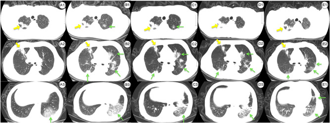
Axial CT images of the chest in patient 3 with the COVID‐19 and tuberculosis coinfection. A, CT obtained on day 2. A1, Stripe of high‐density shadow with a cavitating lesion in the right upper lobe. A2, Stripe of high‐density shadow in the right upper lobe. A3, Pathy ground‐glass opacities (GGOs) in the left lower lobe. B, CT obtained on day 7. B1, Stripe of high‐density shadow with a cavitating lesion in the right upper lobe, slight GGO in the left upper lobe. B2, Stripe of high‐density shadow on the right middle lobe, subpleural (GGOs) in the right upper lobe. Multifocal, limited GGO is seen in the left lungs. B3, Increased multiple GGOs and consolidation in the left lower lobe with air bronchograms. C, CT obtained on day 16. C1, Stripe of high‐density shadow with a cavitating lesion in the right upper lobe, slight GGO in the left upper lobe. C2, Stripe of high‐density shadow on the right middle lobe, absorption of GGO bilaterally. C3, GGO and consolidation in left lower lobe. D, CT obtained on day 22. D1, Stripe of high‐density shadow with a cavitating lesion in the right upper lobe, absorption of GGO in the left upper lobe. D2, Stripe of high‐density shadow on the right middle lobe, absorption of GGO bilaterally. D3, GGO and consolidation in left lower lobe. E, Follow‐up CT obtained on day 59. E1, Stripe of high‐density shadow with a cavitating lesion in the right upper lobe. E2, Stripe of high‐density shadow on right middle lobe, absorption of GGO bilaterally. E3, Absorption of GGO in the left lower lobe. Yellow arrows demonstrated the lesion of tuberculosis. Green arrows demonstrated the lesion of COVID‐19. COVID‐19, coronavirus disease 2019; CT, computed tomography
References
-
- World Health Organization . Statement on the second meeting of the International Health Regulations (2005) Emergency Committee regarding the outbreak of novel coronavirus (2019‐nCoV). https://www.who.int/news-room/detail/30-01-2020-statement-on-the-second-.... Accessed 15 February 2020.
-
- Griffin DE, Bellini WJ. Measles virus. In: Fields BN, Knipe DM, Howley PM, eds. Virology. Philadelphia, PA: Lippincott‐Raven; 1996:1267‐1312.
-
- Kempe CH, Fulginiti VA. The pathogenesis of measles virus infection. Arch Gesamte Virusforsch. 1965;16:103‐128. - PubMed
-
- Kim HY, Song KS, Goo JM, Lee JS, Lee KS, Lim TH. Thoracic sequelae and complications of tuberculosis. Radiographics. 2001;21(4):839‐858. discussion 859‐860. - PubMed
-
- Stepanian I. Bronchial impotence in patients with pulmonary tuberculosis [article in Russian]. Tuberk Biolezni Legk. 2013;4(1):6‐11.
Publication types
MeSH terms
Grants and funding
LinkOut - more resources
Full Text Sources
Medical
