Review
. 2020 Mar 27;117(13):225-234.
doi: 10.3238/arztebl.2020.0225.

The Diagnosis and Treatment of Glaucoma

Affiliations
Review

The Diagnosis and Treatment of Glaucoma

Alexander K Schuster et al. Dtsch Arztebl Int. .

Abstract

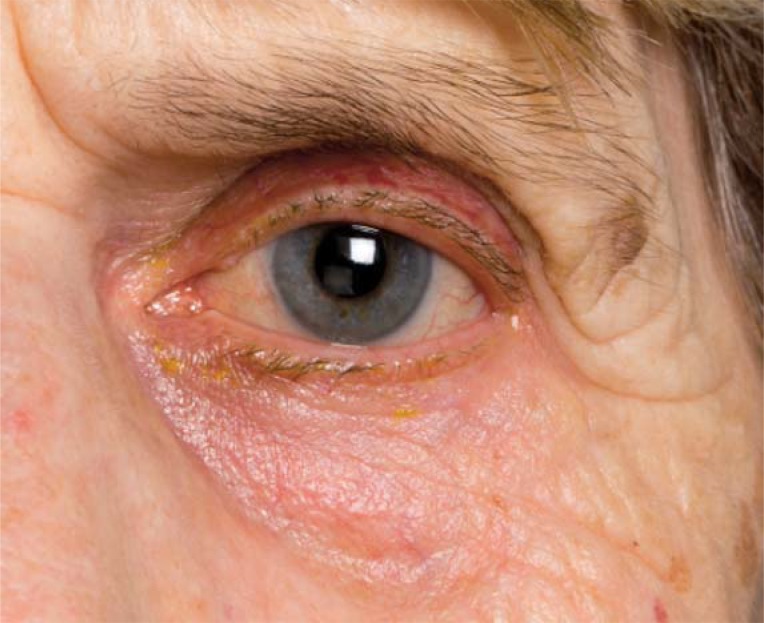
Background: Glaucoma is a group of chronically progressive disorders of the optic nerve. In this article, we present the epidemiology of and risk factors for glaucoma, as well as the diagnostic work-up and treatment options.

Methods: This review is based on pertinent publications retrieved by a selective search in Medline and the Cochrane Library, supplemented by further articles chosen by the authors.

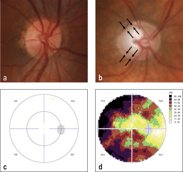
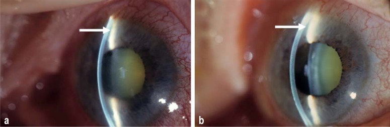
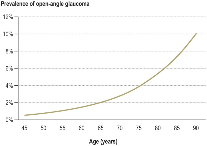
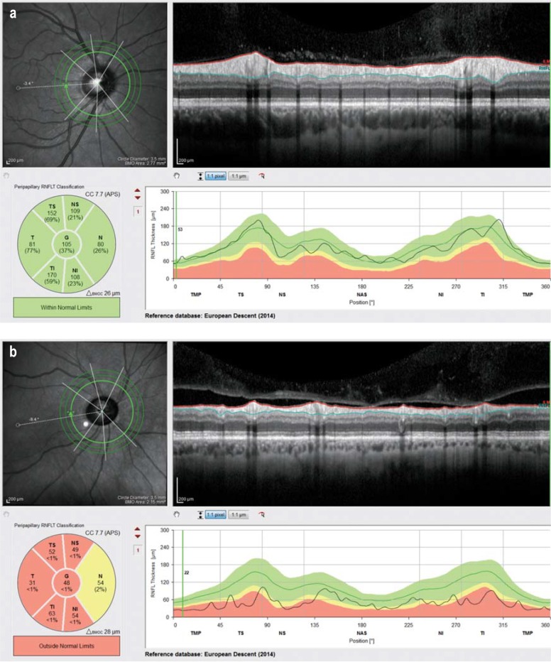
Results: In Europe, the prevalence of glaucoma is 2.93% among persons aged 40 to 80 years. The prevalence rises with age, reaching 10% in persons over 90 years old. The available diagnostic methods include ophthalmoscopy, tonometry, perimetry, and imaging techniques. The treatment of glaucoma is focused on lowering the intraocular pressure with topical drugs, laser therapy, and glaucoma surgery. In patients with manifest glaucoma, lowering the intraocular pressure prevents the progression of visual field defects, with a number needed to treat of 7.

Conclusion: The diagnostic evaluation of glaucoma rests on multiple pillars, all of which must be considered for establishing the diagnosis and defining the desired target pressure: these are, among others, the intraocular pressure and ocular function and morphology. Individually tailored pressure-lowering treatment should be evaluated in regularly scheduled follow-up visits for assessment of function and morphology and adjusted as necessary to minimize the risk of progression.


Figures

Figure 1:
Figure 1:
Optic disc images: a) normal and b) glaucomatous optic disc; c) and d) the respective visual field measurements. The findings from one eye (the right eye) are shown in all images. The optic disc in b) displays, particularly in the upper and lower temporal quadrants, marked thinning of the neuroretinal edge zone (black arrows), with a large excavation. The corresponding visual field examination d) reveals marked defects with central sparing. Darker shading represents the areas in which light is less well perceived; a normal visual field is shown for comparison (c). Visual field measurements depend on the patient’s concentration and cooperation, which can be quantified, for example, with automatic fixation detection and trick questions. Images: Department of Ophthalmology, University Medical Center Mainz
Figure 2
Figure 2
Slit-lamp examination a) of an occluded chamber angle in acute angle closure and b) after successful treatment (laser iridotomy). There is marked enlargement of the chamber angle (white arrow) and deepening of the anterior chamber from a) to b), as well as corneal clearing and regression of conjunctival hyperemia and scleral vascular distention. Images: Department of Ophthalmology, University Medical Center Mainz
Figure 3
Figure 3
The increasing prevalence of glaucoma with advancing age in persons of European extraction. Data from Kapetanakis et al. (19)
Figure 4
Figure 4
Examination of peripapillary nerve fiber layer thickness a) in a normal eye and b) in a glaucomatous eye. The retinal nerve fiber layer lies between the internal limiting membrane (red line) and the border between the retinal nerve fiber layer and the ganglion cell layer (turquoise line). Beyond this layer, reflections from the vitreous body can be seen. Images: Department of Ophthalmology, University Medical Center Mainz
eFigure:
eFigure:
Side effects of local antiglaucomatous treatment There is redness of the periocular skin, as well as a reduction of periorbital fat and mild conjunctival hyperemia. Images: Department of Ophthalmology, University Medical Center Mainz

References

    1. Leffler CT, Schwartz SG, Giliberti FM, et al. What was glaucoma called before the 20th century? Ophthalmol Eye Dis. 2015;7:21–33. - PMC - PubMed
    1. Weinreb RN, Aung T, Medeiros FA. The pathophysiology and treatment of glaucoma: a review. JAMA. 2014;311:1901–1911. - PMC - PubMed
    1. Jonas JB, Aung T, Bourne RR, et al. Glaucoma. Lancet. 2017;390:2183–2193. - PubMed
    1. Tian H, Li L, Song F. Study on the deformations of the lamina cribrosa during glaucoma. Acta Biomater. 2017;55:340–348. - PubMed
    1. Shoji T, Kuroda H, Suzuki M, et al. Glaucomatous changes in lamina pores shape within the lamina cribrosa using wide bandwidth, femtosecond mode-locked laser OCT. PLoS One. 2017;12 e0181675. - PMC - PubMed