. 2020 Jun 4;5(11):e137189.
doi: 10.1172/jci.insight.137189.

Cholesterol 25-hydroxylase promotes efferocytosis and resolution of lung inflammation

Affiliations

Cholesterol 25-hydroxylase promotes efferocytosis and resolution of lung inflammation

Jennifer H Madenspacher et al. JCI Insight. .

Abstract

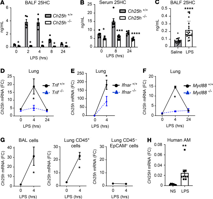
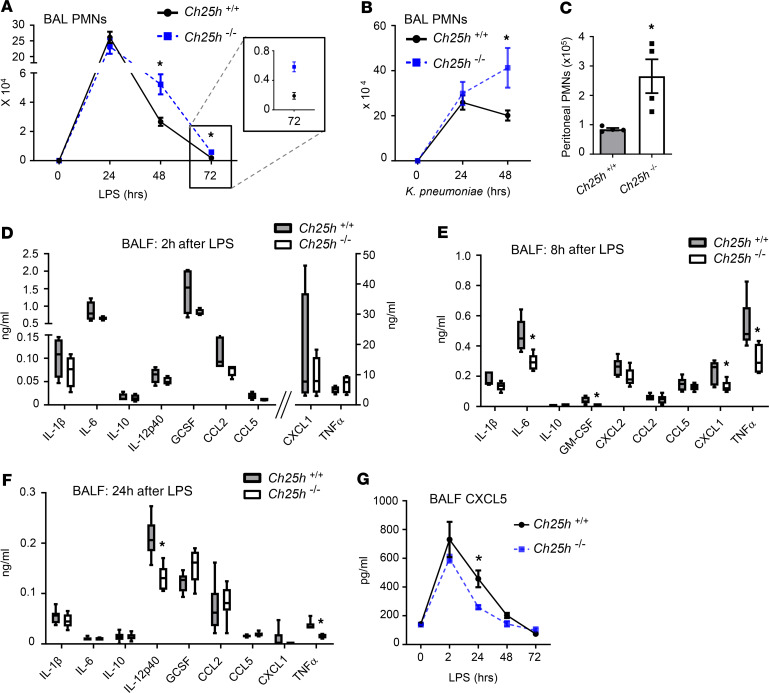
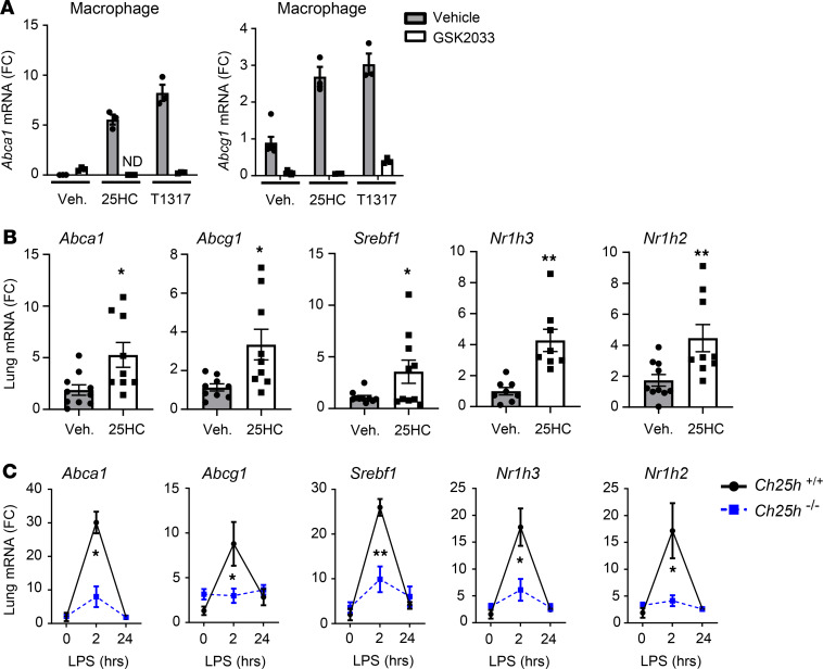
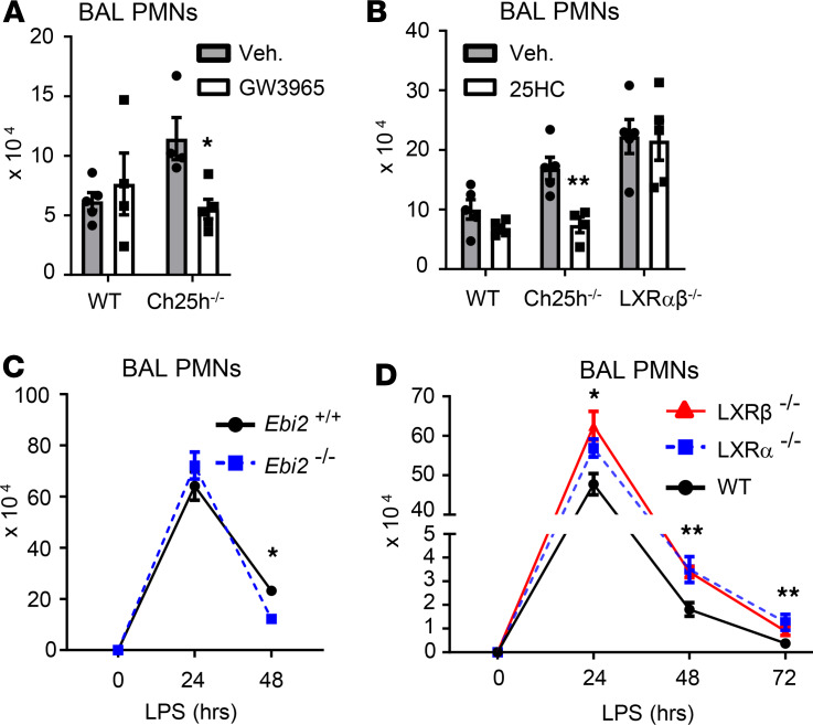
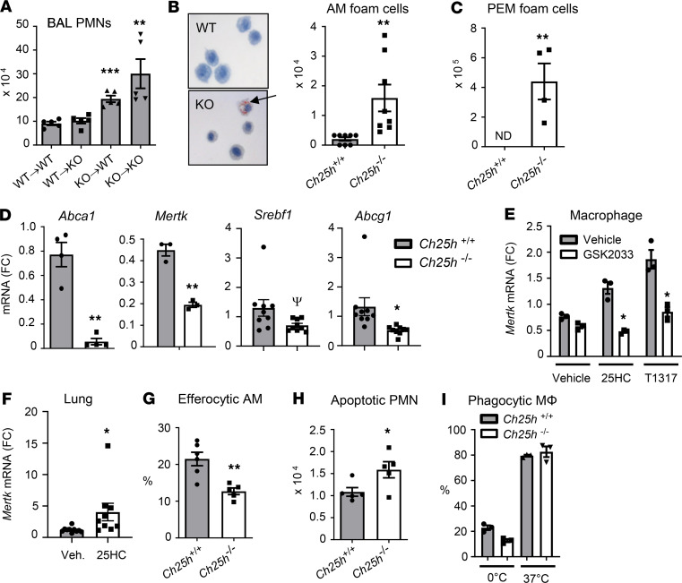
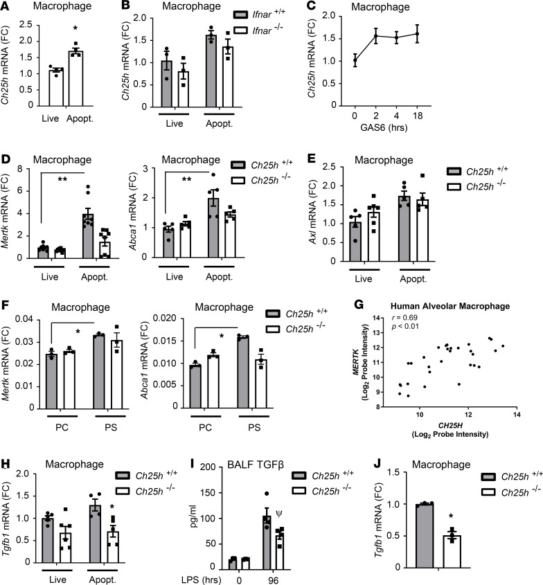
Alveolar macrophages (AM) play a central role in initiation and resolution of lung inflammation, but the integration of these opposing core functions is poorly understood. AM expression of cholesterol 25-hydroxylase (CH25H), the primary biosynthetic enzyme for 25-hydroxycholesterol (25HC), far exceeds the expression of macrophages in other tissues, but no role for CH25H has been defined in lung biology. As 25HC is an agonist for the antiinflammatory nuclear receptor, liver X receptor (LXR), we speculated that CH25H might regulate inflammatory homeostasis in the lung. Here, we show that, of natural oxysterols or sterols, 25HC is induced in the inflamed lung of mice and humans. Ch25h-/- mice fail to induce 25HC and LXR target genes in the lung after LPS inhalation and exhibit delayed resolution of airway neutrophilia, which can be rescued by systemic treatment with either 25HC or synthetic LXR agonists. LXR-null mice also display delayed resolution, suggesting that native oxysterols promote resolution. During resolution, Ch25h is induced in macrophages upon their encounter with apoptotic cells and is required for LXR-dependent prevention of AM lipid overload, induction of Mertk, efferocytic resolution of airway neutrophilia, and induction of TGF-β. CH25H/25HC/LXR is, thus, an inducible metabolic axis that programs AMs for efferocytic resolution of inflammation.

Keywords: Cholesterol; Inflammation; Innate immunity; Macrophages; Pulmonology.


Conflict of interest statement

Conflict of interest: The authors have declared that no conflict of interest exists.

Figures

Figure 1
Figure 1. Ch25h is induced by LPS in macrophages of murine and human lung.
(A and B) BALF (A) and serum 25HC (B) was quantified by liquid chromatography–mass spectrometry (LC-MS) at the indicated time points following LPS inhalation in Ch25h+/+ and Ch25h–/– mice (n = 5–7/genotype/time point). (C) BALF 25HC was quantified 16 hours following contralateral subsegmental bronchoscopic instillation of saline and LPS into human volunteers (n = 23) (D–F) Lung tissue Ch25h mRNA (fold change [FC], normalized to Gapdh and indexed to WT/control) was quantified by quantitative PCR (qPCR) at various times after LPS inhalation in the murine strains indicated (n = 5/genotype/time point). (G) Ch25h mRNA (Gapdh-normalized) was quantified in BAL cells, and in CD45+ and CD45EpCAM+ cells from WT lung parenchyma at the indicated times in relation to LPS inhalation (n = 3/genotype/time point). (H) Alveolar macrophages (AM) collected from BAL of normal, healthy human controls (n = 3) were exposed ex vivo to LPS (10 ng/mL, 4 hours) or left nonstimulated (NS) and then assayed by qPCR for normalized CH25H mRNA. Data are mean ± SEM and are representative of 2–3 independent experiments. *P < 0.05; **P < 0.01; ****P < 0.0001 by unpaired t test.
Figure 2
Figure 2. CH25H is required for resolution of neutrophilia.
(A and B) Ch25h+/+ and Ch25h–/– mice were exposed to LPS (A) or K. pneumoniae (B) by inhalation and then BAL polymorphonuclear neutrophils (PMNs) were quantified at the indicated times (n = 6/genotype/time point). (C) Peritoneal PMNs were quantified in Ch25h+/+ and Ch25h–/– mice 4 days following i.p. injection of Brewer’s thioglycollate (n = 4/genotype). (D–F) BALF was collected from Ch25h+/+ and Ch25h–/– mice (n = 4–6/genotype/time point) at 2 hours (D), 8 hours (E), and 24 hours (F) after inhalation of LPS and analyzed for cytokine levels by multiplex assay. (G) BALF was collected from mice (n = 3–5/genotype/time point) at the indicated times following inhaled LPS and analyzed by ELISA for CXCL5 concentration. Data are mean ± SEM and are representative of 2–3 independent experiments. *P < 0.05 by unpaired t test.
Figure 3
Figure 3. 25HC activates LXR in the lung.
(A) WT peritoneal exudate macrophages (PEMs) were exposed for 16 hours to 25HC or LXR agonist TO901317 (T1317) in the presence or absence of the LXR antagonist GSK2033 and were then quantified by qPCR for Abca1 or Abcg1 mRNA (fold change [FC], n = 3/condition; ND, not detected). (B) WT mice were treated i.p. with 25HC or vehicle (Veh.), and then lung tissue was analyzed by qPCR for the targets shown (n = 8–10/condition). (C) Ch25h+/+ and Ch25h–/– mice were exposed to inhaled LPS, and lung tissue was analyzed by qPCR for the indicated targets at the times shown (n = 4–6/genotype/time point). Data are mean ± SEM and are representative of 2–3 independent experiments. *P < 0.05; **P < 0.01 by unpaired t test.
Figure 4
Figure 4. Defective resolution in Ch25h-null mice arises from LXR insufficiency.
(A and B) Mice of the indicated genotypes were pretreated i.p. with either synthetic LXR agonist GW3965 (A) or 25HC (B) (or vehicle [Veh] control), exposed to inhaled LPS, and then evaluated for airway PMN count 48 hours later (n = 5/genotype/time point). (C and D) Airway PMNs were quantified in mice of the indicated genotypes at various times following inhalation of LPS (n = 5–8/genotype/time point). Data are mean ± SEM and are representative of 2–3 independent experiments. *P < 0.05; **P < 0.01 by unpaired t test.
Figure 5
Figure 5. Ch25h-null macrophages display cholesterol dysregulation and defective efferocytosis.
(A) Chimeric mice generated by bone marrow transfer from Ch25h+/+ (WT) or Ch25h–/– (KO) donors to irradiated WT or KO recipients (shown as donor → recipient) were exposed to inhaled LPS, and bronchoalveolar lavage (BAL) PMNs were quantified 48 hours later (n = 5/genotype/time point). (B) Alveolar macrophages (AMs) from Ch25h+/+ and Ch25h–/– mice collected 48 hours after LPS inhalation were stained with Oil Red O (left) and stain-positive (foam) cells quantified (right) (n = 8/genotype). Arrow indicates a stain-positive cell. (C) A similar analysis as shown in B was conducted on peritoneal exudate macrophages (PEMs) harvested 72 hours after i.p. thioglycollate (n = 4/genotype). (D) PEMs freshly harvested from Ch25h+/+ and Ch25h–/– mice (n = 3–9/genotype) were analyzed by qPCR for the targets shown (fold change, FC). (E) WT PEMs were treated with 25HC or T0901317 in the presence or absence of LXR antagonist GSK2033 and then analyzed by qPCR for normalized Mertk mRNA. (F) WT mice were treated i.p. with 25HC or vehicle (Veh.), and then lung tissue was analyzed by qPCR for Mertk (n = 8–10/condition). (G) Ch25h+/+ and Ch25h–/– mice (n = 6/genotype) were instilled i.t. with apoptotic WT thymocytes 48 hours after LPS inhalation. AMs that had internalized thymocytes (efferocytic AMs) were manually quantified 90 minutes later. (H) Apoptotic (FLIVO+) PMNs were quantified in BALF of Ch25h+/+ and Ch25h–/– mice (n = 5/genotype) 48 hours after LPS inhalation. (I) Internalization of bacterial bioparticles by Ch25h+/+ and Ch25h–/– PEMs was quantified at 37°C and 0°C (control). Data are mean ± SEM and are representative of 3–6 independent experiments. ΨP = 0.06; *P < 0.05; **P < 0.01; ***P < 0.001 by unpaired t test.
Figure 6
Figure 6. Apoptotic cells induce Ch25h-dependent Mertk in efferocytic macrophages.
(A) WT peritoneal exudate macrophages (PEMs) were cocultured for 16 hours with live or apoptotic Jurkat T cells, after which Ch25h mRNA was quantified by qPCR (fold change [FC], n = 4/condition). (B) Ifnar+/+ and Ifnar–/– PEMs were treated and analyzed as in A (n = 3/condition). (C) WT PEMs (n = 4/condition) were treated with 400 ng/mL Gas6 protein for the indicated times and then analyzed by qPCR for Ch25h mRNA. (D and E) Ch25h+/+ and Ch25h–/– PEMs were treated as in B and then analyzed by qPCR for the indicated targets (n = 5–8/condition). (F) Ch25h+/+ and Ch25h–/– PEMs were treated with phosphatidylserine (PS) or phosphatidylcholine (PC [control]) liposomes (400 μg/mL, 4 hours) and then analyzed by qPCR for the indicated targets (n = 3/condition). (G) RNA purified from AMs isolated by negative selection from patients (n = 30) enrolled within 48 hours after ARDS onset in the omega-3 fatty acids trial (50) was analyzed by microarray. A Pearson’s test was used to generate a correlation coefficient between normalized log2 CH25H and MERTK probe intensities. (H) Ch25h+/+ and Ch25h–/– PEMs were treated as in D and then analyzed by qPCR for Tgfb1 mRNA (n = 4–6/condition). (I) Mice of the indicated genotypes were exposed to 3 mg/mL LPS aerosol and BALF TGF-β analyzed by ELISA (n = 3–4/condition). (J) PEMs freshly harvested from Ch25h+/+ and Ch25h–/– mice were analyzed by qPCR for Tgfb1 mRNA (n = 3/genotype). Data are mean ± SEM and are representative of 2–4 independent experiments. *P < 0.05; **P < 0.01; ψP = 0.059 by unpaired t test other than for G.

References

    1. Mould KJ, et al. Cell Origin Dictates Programming of Resident versus Recruited Macrophages during Acute Lung Injury. Am J Respir Cell Mol Biol. 2017;57(3):294–306. doi: 10.1165/rcmb.2017-0061OC. - DOI - PMC - PubMed
    1. Janssen WJ, et al. Surfactant proteins A and D suppress alveolar macrophage phagocytosis via interaction with SIRP alpha. Am J Respir Crit Care Med. 2008;178(2):158–167. doi: 10.1164/rccm.200711-1661OC. - DOI - PMC - PubMed
    1. Mohning MP, et al. Phagocytosis of microparticles by alveolar macrophages during acute lung injury requires MerTK. Am J Physiol Lung Cell Mol Physiol. 2018;314(1):L69–L82. doi: 10.1152/ajplung.00058.2017. - DOI - PMC - PubMed
    1. Huynh ML, Fadok VA, Henson PM. Phosphatidylserine-dependent ingestion of apoptotic cells promotes TGF-beta1 secretion and the resolution of inflammation. J Clin Invest. 2002;109(1):41–50. doi: 10.1172/JCI11638. - DOI - PMC - PubMed
    1. Fadok VA, Bratton DL, Konowal A, Freed PW, Westcott JY, Henson PM. Macrophages that have ingested apoptotic cells in vitro inhibit proinflammatory cytokine production through autocrine/paracrine mechanisms involving TGF-beta, PGE2, and PAF. J Clin Invest. 1998;101(4):890–898. doi: 10.1172/JCI1112. - DOI - PMC - PubMed

Publication types