

Circulating cell free DNA during definitive chemo-radiotherapy in non-small cell lung cancer patients - initial observations
- PMID: 32343749
- PMCID: PMC7188247
- DOI: 10.1371/journal.pone.0231884
Circulating cell free DNA during definitive chemo-radiotherapy in non-small cell lung cancer patients - initial observations
Abstract
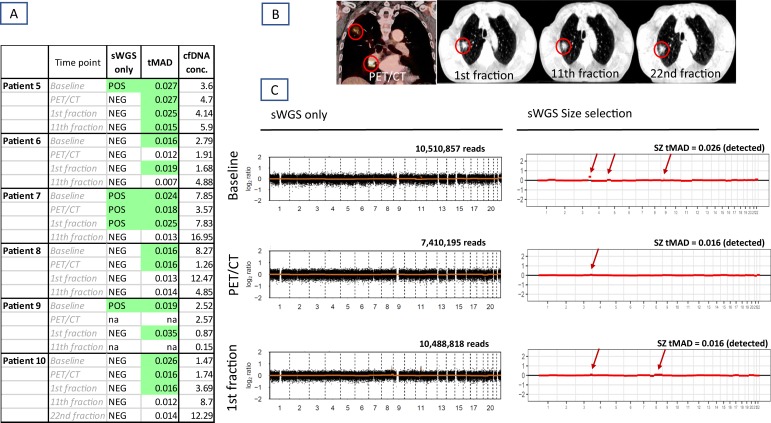
Background: The overall aim was to investigate the change over time in circulating cell free DNA (cfDNA) in patients with locally advanced non-small cell lung cancer (NSCLC) undergoing concurrent chemo-radiotherapy. Furthermore, to assess the possibility of detecting circulating cell free tumor DNA (ctDNA) using shallow whole genome sequencing (sWGS) and size selection.
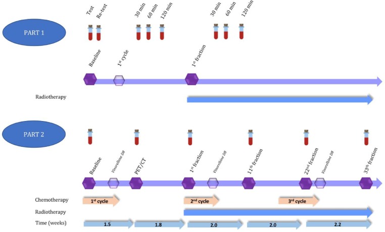
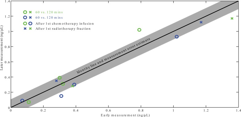
Methods: Ten patients were included in a two-phase study. The first four patients had blood samples taken prior to a radiation therapy (RT) dose fraction and at 30 minutes, 1 hour and 2 hours after RT to estimate the short-term dynamics of cfDNA concentration after irradiation. The remaining six patients had one blood sample taken on six treatment days 30 minutes post treatment to measure cfDNA levels. Presence of ctDNA as indicated by chromosomal aberrations was investigated using sWGS. The sensitivity of this method was further enhanced using in silico size selection.
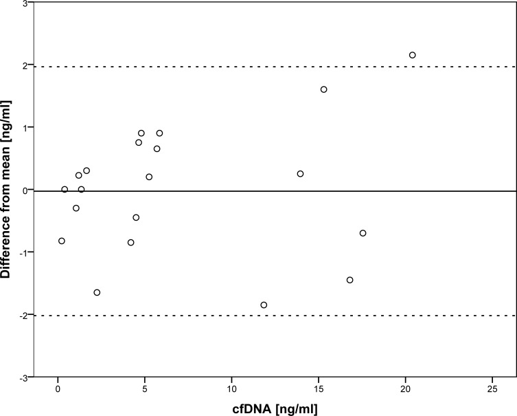
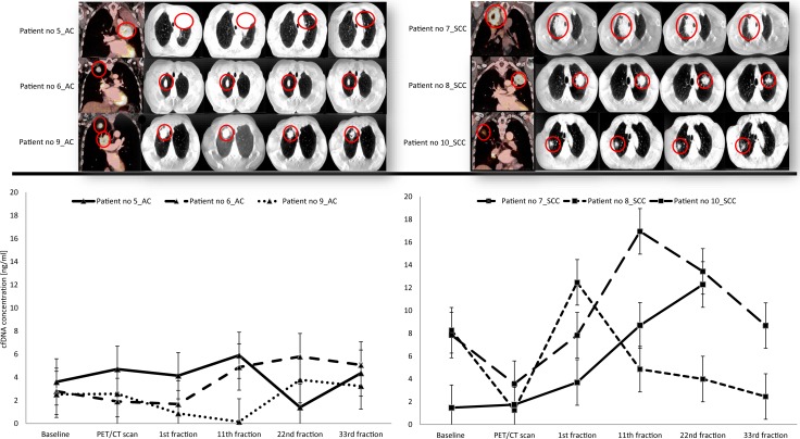
Results: cfDNA concentration from baseline to 120 min after therapy was stable within 95% tolerance limits of +/- 2 ng/ml cfDNA. Changes in cfDNA were observed during therapy with an apparent qualitative difference between adenocarcinoma (average increase of 0.69 ng/ml) and squamous cell carcinoma (average increase of 4.0 ng/ml). Tumor shrinkage on daily cone beam computer tomography scans during radiotherapy did not correlate with changes in concentration of cfDNA.
Conclusion: Concentrations of cfDNA remain stable during the first 2 hours after an RT fraction. However, based on the sWGS profiles, ctDNA represented only a minor fraction of cfDNA in this group of patients. The detection sensitivity of genomic alterations in ctDNA strongly increases by applying size selection.
Conflict of interest statement
The authors have declared that no competing interests exist.
Figures
References
-
- Dehing-Oberije C, De Ruysscher D, van der Weide H, Hochstenbag M, Bootsma G, Geraedts W, et al. Tumor volume combined with number of positive lymph node stations is a more important prognostic factor than TNM stage for survival of non-small-cell lung cancer patients treated with (chemo)radiotherapy. International journal of radiation oncology, biology, physics. 2008. March 15;70(4):1039–44. 10.1016/j.ijrobp.2007.07.2323 - DOI - PubMed
-
- Berghmans T, Dusart M, Paesmans M, Hossein-Foucher C, Buvat I, Castaigne C, et al. Primary tumor standardized uptake value (SUVmax) measured on fluorodeoxyglucose positron emission tomography (FDG-PET) is of prognostic value for survival in non-small cell lung cancer (NSCLC): a systematic review and meta-analysis (MA) by the European Lu. Journal of thoracic oncology: official publication of the International Association for the Study of Lung Cancer. 2008. January;3(1):6–12. - PubMed
-
- Merker JD, Oxnard GR, Compton C, Diehn M, Hurley P, Lazar AJ, et al. JOURNAL OF CLINICAL ONCOLOGY Circulating Tumor DNA Analysis in Patients With Cancer: American Society of Clinical Oncology and College of American Pathologists Joint Review. J Clin Oncol. 2018;36:1631–41. 10.1200/JCO.2017.76.8671 - DOI - PubMed
-
- Van Der Vaart M, Pretorius PJ. Circulating DNA: Its origin and fluctuation. Annals of the New York Academy of Sciences. 2008;1137(0):18–26. - PubMed
Publication types
MeSH terms
Substances
Grants and funding
LinkOut - more resources
Full Text Sources
Medical
