

Porous Silicon-Zinc Oxide Nanocomposites Prepared by Atomic Layer Deposition for Biophotonic Applications
- PMID: 32344562
- PMCID: PMC7216101
- DOI: 10.3390/ma13081987
Porous Silicon-Zinc Oxide Nanocomposites Prepared by Atomic Layer Deposition for Biophotonic Applications
Abstract
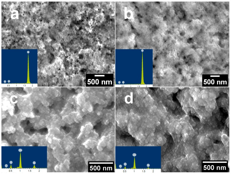
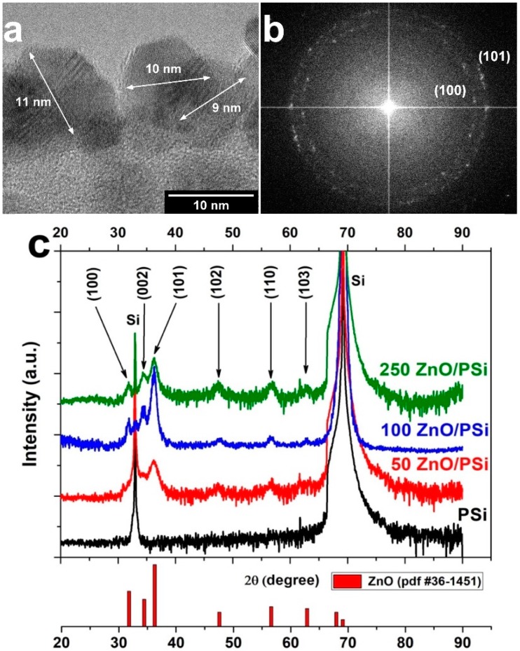
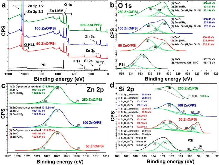
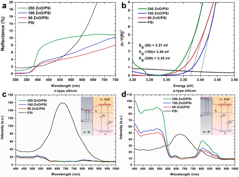
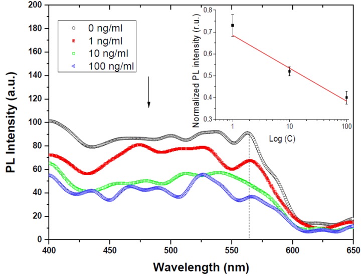
In the current research, a porous silicon/zinc oxide (PSi/ZnO) nanocomposite produced by a combination of metal-assisted chemical etching (MACE) and atomic layer deposition (ALD) methods is presented. The applicability of the composite for biophotonics (optical biosensing) was investigated. To characterize the structural and optical properties of the produced PSi/ZnO nanocomposites, several studies were performed: scanning and transmission electron microscopy (SEM/TEM), X-ray diffraction (XRD), X-ray photoelectron spectroscopy (XPS), diffuse reflectance, and photoluminescence (PL). It was found that the ALD ZnO layer fully covers the PSi, and it possesses a polycrystalline wurtzite structure. The effect of the number of ALD cycles and the type of Si doping on the optical properties of nanocomposites was determined. PL measurements showed a "shoulder-shape" emission in the visible range. The mechanisms of the observed PL were discussed. It was demonstrated that the improved PL performance of the PSi/ZnO nanocomposites could be used for implementation in optical biosensor applications. Furthermore, the produced PSi/ZnO nanocomposite was tested for optical/PL biosensing towards mycotoxins (Aflatoxin B1) detection, confirming the applicability of the nanocomposites.
Keywords: atomic layer deposition; biosensors; photoluminescence; porous silicon; zinc oxide.
Conflict of interest statement
The authors declare no conflict of interest.
Figures
References
-
- Iatsunskyi I., Smyntyna V., Pavlenko M., Kanevska O., Kirik Y., Myndrul V. Ammonia detection using optical reflectance from porous silicon formed by metal-assisted chemical etching; Proceedings of the Optics and Photonics for Counterterrorism, Crime Fighting and Defence IX; and Optical Materials and Biomaterials in Security and Defence Systems Technology X; Dresden, Germany. 23–24 September 2013; p. 8901.
-
- Tischler M.A., Collins R.T., Stathis J.H., Tsang J.C. Luminescence degradation in porous silicon. Appl. Phys. Lett. 1992;60:639–641. doi: 10.1063/1.106578. - DOI
-
- Iatsunskyi I., Jancelewicz M., Nowaczyk G., Kempiński M., Peplińska B., Jarek M., Załęski K., Jurga S., Smyntyna V. Atomic layer deposition TiO2 coated porous silicon surface: Structural characterization and morphological features. Thin Solid Films. 2015;589:303–308. doi: 10.1016/j.tsf.2015.05.056. - DOI
-
- Brytavskyi I., Hušeková K., Myndrul V., Pavlenko M., Coy E., Zaleski K., Gregušová D., Yate L., Smyntyna V., Iatsunskyi I. Effect of porous silicon substrate on structural, mechanical and optical properties of MOCVD and ALD ruthenium oxide nanolayers. Appl. Surf. Sci. 2019;471:686–693. doi: 10.1016/j.apsusc.2018.12.022. - DOI
Grants and funding
LinkOut - more resources
Full Text Sources
