

Digitization of One-Piece Oral Implants: A Feasibility Study
- PMID: 32344639
- PMCID: PMC7215390
- DOI: 10.3390/ma13081990
Digitization of One-Piece Oral Implants: A Feasibility Study
Abstract
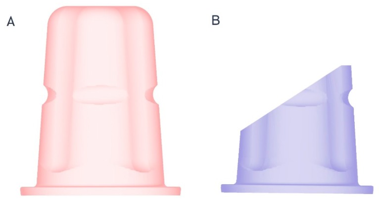
For digital impression-making of two-piece oral implants, scan bodies are used to transfer the exact intraoral implant position to the dental laboratory. In this in vitro investigation, the accuracy of digitizing a one-piece ceramic oral implant without a scan body (OC) was compared to that of a standard two-piece titanium implant with a scan body (TT) and a preparation of a natural single tooth (ST). Furthermore, incomplete scans of OC simulating clinical compromising situations (OC1-4) were redesigned using a virtual reconstruction tool (RT) and superimposed to OC. OC and TT oral implants and one ST were inserted into a mandible typodont model and digitized (N = 13) using two different intraoral scanners. The resulting virtual datasets were superimposed onto a three-dimensional (3D) laser scanner-based reference. Test and reference groups were aligned using an inspection software according to a best-fit algorithm, and circumferential as well as marginal discrepancies were measured. For the statistical evaluation, multivariate analyses of variance with post-hoc Tukey tests and students t-tests to compare both scanners were performed. A total of 182 datasets were analyzed. For circumferential deviations, no significant differences were found between ST, TT, and OC (p > 0.964), but increased deviations for OC1-4 (p < 0.001) were observed. The measurements of the marginal deviations revealed that ST had the smallest deviations, and that there were no significant differences between TT, OC, and OC1-4 (p > 0.979). Except for marginal deviation of OC (p < 0.001), the outcome was not affected by the scanner. Within the limitations of this study, digitization of OC is as accurate as that of TT, but less than that of ST. In the case of known geometries, post-processing of compromised scans with a virtual reconstruction results in accurate data.
Keywords: accuracy; ceramics; dental implants; digital impression; intraoral scanner; one-piece dental implant; trueness.
Conflict of interest statement
The authors declare no conflicts of interest. The funders had no role in the design of the study; in the collection, analyses, or interpretation of data; in the writing of the manuscript; or in the decision to publish the results.
Figures


References
-
- Su T.S., Sun J. Intraoral Digital Impression Technique: A Review. J. Prosthodont. 2015;24:313–321. - PubMed
Grants and funding
LinkOut - more resources
Full Text Sources
