. 2020 Apr 24;9(4):1062.
doi: 10.3390/cells9041062.

Inhibiting Monocyte Recruitment to Prevent the Pro-Tumoral Activity of Tumor-Associated Macrophages in Chondrosarcoma

Affiliations

Inhibiting Monocyte Recruitment to Prevent the Pro-Tumoral Activity of Tumor-Associated Macrophages in Chondrosarcoma

Michele Minopoli et al. Cells. .

Abstract

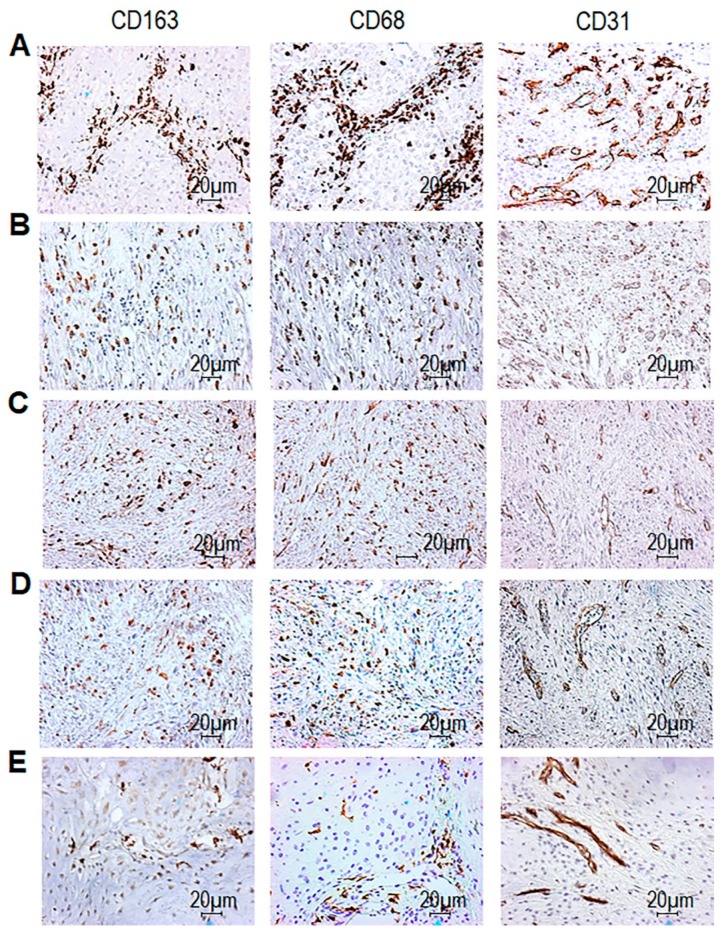
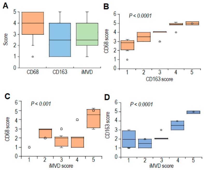
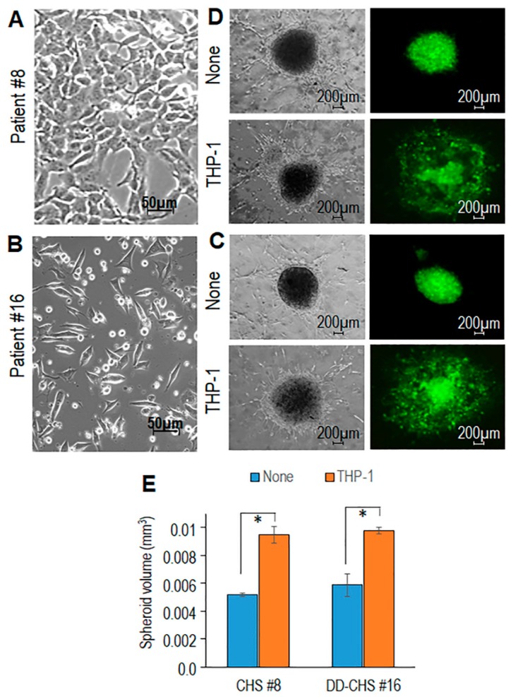
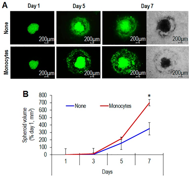
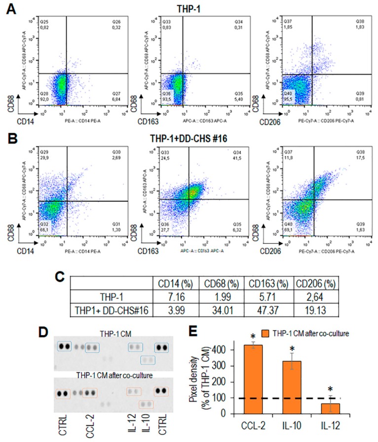
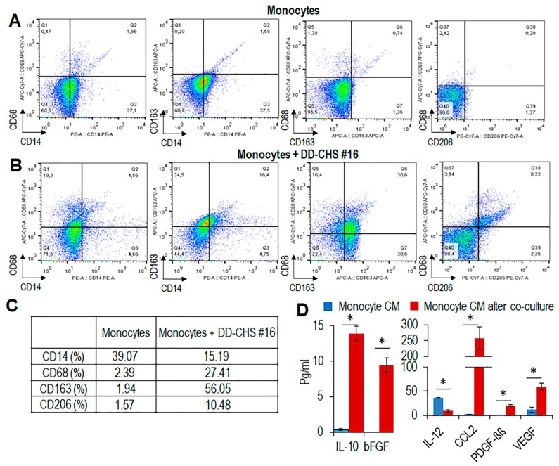
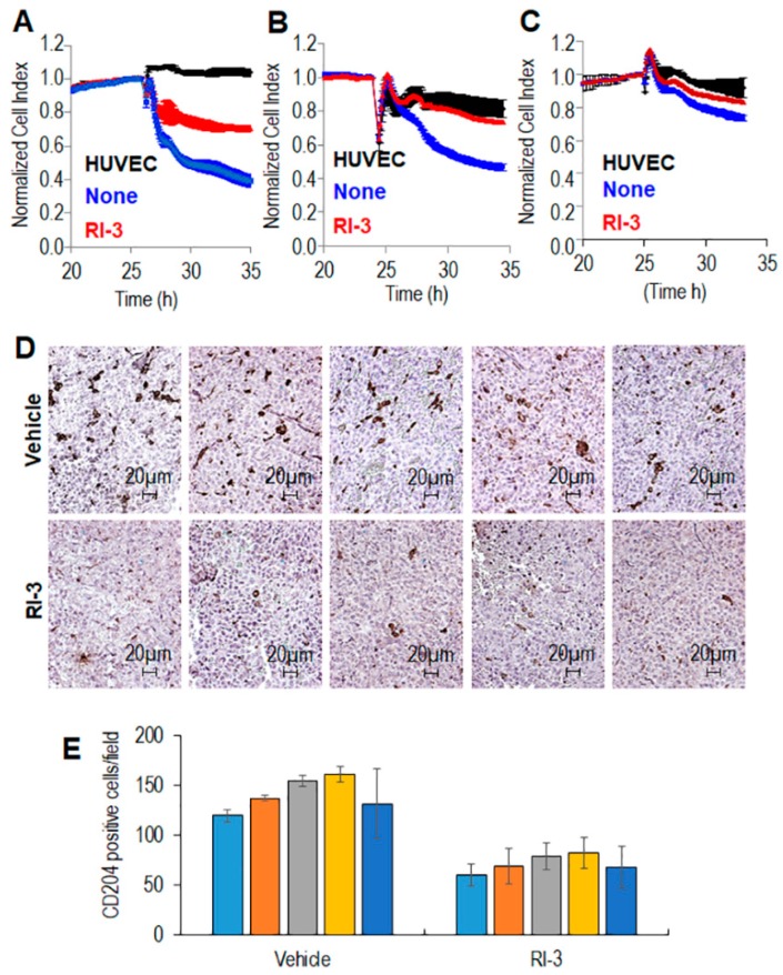
Chondrosarcomas (CHS) are malignant cartilaginous neoplasms with diverse morphological features, characterized by resistance to chemo- and radiation therapies. In this study, we investigated the role of tumor-associated macrophages (TAM)s in tumor tissues from CHS patients by immunohistochemistry. Three-dimensional organotypic co-cultures were set up in order to evaluate the contribution of primary human CHS cells in driving an M2-like phenotype in monocyte-derived primary macrophages, and the capability of macrophages to promote growth and/or invasiveness of CHS cells. Finally, with an in vivo model of primary CHS cells engrafted in nude mice, we tested the ability of a potent peptide inhibitor of cell migration (Ac-d-Tyr-d-Arg-Aib-d-Arg-NH2, denoted RI-3) to reduce recruitment and infiltration of monocytes into CHS neoplastic lesions. We found a significant correlation between alternatively activated M2 macrophages and intratumor microvessel density in both conventional and dedifferentiated CHS human tissues, suggesting a link between TAM abundance and vascularization in CHS. In 3D and non-contact cu-culture models, soluble factors produced by CHS induced a M2-like phenotype in macrophages that, in turn, increased motility, invasion and matrix spreading of CHS cells. Finally, we present evidence that RI-3 successfully prevent both recruitment and infiltration of monocytes into CHS tissues, in nude mice.

Keywords: cell migration inhibitors; chondrosarcoma; monocytes; tumor-associated macrophages.


Conflict of interest statement

The authors declare no conflict of interest.

Figures

Figure 1
Figure 1
CD68, CD163, and CD31 expression in tumor tissues from chondrosarcomas (CHS) patients. CHS tissues were processed for IHC analysis of CD68+ and CD163+ cells, and CD31+ microvessels. Sections were scored based on the average counts of positive cells (CD68 and CD163) or microvessels (CD31) counted in the tumor areas, in five randomly selected fields/sample at 200× magnification. For each section, positive cells or microvessels were scored as 1-5 where 1 (1–25), 2 (26–50), 3 (51–100), 4 (101–150), and 5 (> 150) positive cells or microvessels were encountered in the field. Representative images of CD163 score 5 (A, from patient #8) score 4 (B, from patient #16) score 3 (C, from patient #14) score 2 (D, from patient #13) and score 1 (E, from patient #10). The corresponding CD68 and CD31 immunostaining are shown in the central and right columns (scoring is reported in the Table 2). Original magnification: 200×. Distribution of CD68, CD163 and i intratumoral microvessel density (iMVD) scores are reported in Table 2 and summarized in Figure 2A.
Figure 2
Figure 2
Relationships between CD68, CD163, and CD31 expression levels in tumor tissues from CHS patients. (A). Box plot, showing variation in the distribution of CD68, CD163 and intratumor microvessel density (iMVD) scores in CHS tissues. (B,C). Box plots showing CD68 scores in CHS tissues according to CD163 (B) and iMVD (C) scores. (D). Box plot, showing scores of CD163 scores in CHS tissues according to iMVD scores. Dark horizontal lines represent the medians. Circles represent outliers.
Figure 3
Figure 3
Contribution of THP-1 cells in promoting spreading of primary CHS cells in organotypic co-cultures. Primary CHS cells obtained from the tumor samples of #8 (A) and #16 (B) patients visualized by phase contrast microscopy. Original magnification: 200x. (C,D). Spheroids of GFP-tagged CHS cells (C: patient #8, D: patient #16) were embedded in a collagen/fibroblast mixture, without (None), or with the addition of THP1 cells. Fluorescent and transmitted-light input images were acquired after 7 days at 50× magnification. (E). Spheroid sizes assessed after 7 days by using the Equation (1), where D and d are the major and the minor diameter, respectively. Data are the mean ± SD of two independent experiments, performed in duplicate. Statistical significance with * p  <  0.0001.
Figure 4
Figure 4
Time-dependent increase of spheroid size induced by primary monocytes. (A). Spheroids containing GFP-tagged CHS cells obtained from the tumor sample of #16 patient were embedded in the collagen/fibroblast mixture without (None), or with the addition of human monocytes. At the indicated times, fluorescent and transmitted-light input images were acquired at 50× magnification. (B). Time-dependent increase of spheroid size. Data expressed as percentage of volumes assessed at time zero are the mean ± SD of two independent experiments, performed in duplicate. Statistical significance with * p < 0.0001.
Figure 5
Figure 5
Immunophenotyping of THP-1 cells co-cultured with CHS cells. THP-1 cells were co-cultured with primary CHS cells for 72 h. (A,B). Phenotypic analysis of THP-1 cells, collected after co-culture, by flow cytometry. (C). Percent variation of CD14+, CD68+, CD163+, and CD206+ cells upon co-culture with CHS cells, compared to control (THP-1 alone). (D). After co-culture, CHS cells were removed, THP-1 conditioned media prepared as described and analyzed for the content of CC2, IL-10 and IL-12 by a dot plot assay. (E). The pixel density of each spot was measured using NIH Image J 2.0 software. Positive control spots were used to normalize results between the membranes. The intensity of each spot was averaged over the duplicate spots and expressed as percentage of each cytokine or chemokine spontaneously secreted by THP-1 cells, considered as 100% (dashed line). Data represent mean ± SD from three experiments performed in quadruplicate with * p < 0.005.
Figure 6
Figure 6
Immunophenotyping of primary human monocytes co-cultured with CHS cells. Human monocytes from healthy donors were co-cultured with primary CHS cells. (A,B). After 72 h, monocytes were recovered and their phenotype analyzed by flow cytometry. (C). Percent variation of CD14+, CD68+ CD163+, and CD206+ cells upon co-culture with CHS cells, compared to control (monocytes alone). (D). After co-culture, CHS were removed, and monocyte conditioned media were prepared and analyzed by a Bio-Plex immunoassay. The concentration of significant soluble factors (expressed in pg/mL) are reported as mean ± SD from two experiments performed in quadruplicate. Statistical significance with * p < 0.001.
Figure 7
Figure 7
Effect of the peptide RI-3 on monocyte recruitment and TAM infiltration into CHS tissues. Endothelial cells (HUVEC) were seeded in E-plates and allowed to adhere for ~25 h, until they formed a confluent monolayer. Then, monolayers were overlaid with THP-1 cells (A), human (B) or murine (C) monocytes in growth medium, in the absence or presence of 10 nM RI-3. Endothelial invasion by monocytes was monitored in real-time for 10 h as changes in Cell Index. Values were normalized immediately after monocyte addition. Data represent mean ± SD from a quadruplicate experiment. (D,E). Ten 6–8 week-old Foxn1nu/nu mice received an injection of CHS cells into the right flank as a single-cell suspension. Animals were randomized into two 5-mice groups with the treatment group receiving 6 mg/kg RI-3 by intra-peritoneal injection every 24 h, and the control group receiving an equivalent injected volume of vehicle. After 12 days, the animals were sacrificed, the excised tumors fixed in buffered formalin, processed for paraffin sectioning and then immunostained with anti-mouse-CD204. (D). Representative images of CD204 immunostaining are shown. (E). CD204+ cells revealed by IHC were counted in 5 randomly chosen fields per section, at 200× magnification and averages plotted. Statistical significance with * p < 0.0001.

References

    1. Hogendoorn P.C.W., Bovée J.V.M.G., Nielsen G.P. WHO Classification of Tumours of Soft Tissue and Bone. 4th ed. HWO Press; Geneva, Switzerland: 2013. pp. 264–268.
    1. Gelderblom H., Hogendoorn P.C.W., Dijkstra S.D., van Rijswijk C.S., Krol A.D., Taminiau A.H.M., Bovée J.V.M.G. The clinical approach towards chondrosarcoma. Oncologist. 2008;13:320–329. doi: 10.1634/theoncologist.2007-0237. - DOI - PubMed
    1. van Maldegem A., Conley A.P., Rutkowski P., Patel S.R., Lugowska I., Desar I.M.E., Bovée J.V.M.G., Gelderblom H. Outcome of First-Line Systemic Treatment for Unresectable Conventional, Dedifferentiated, Mesenchymal, and Clear Cell Chondrosarcoma. Oncologist. 2019;24:110–116. doi: 10.1634/theoncologist.2017-0574. - DOI - PMC - PubMed
    1. Miao R., Choy E., Raskin K.A., Schwab J.H., Nielsen G.P., Deshpande V., Chebib I., DeLaney T.F., Hornicek F.J., Cote G.M., et al. Prognostic Factors in Dedifferentiated Chondrosarcoma: A Retrospective Analysis of a Large Series Treated at a Single Institution. Sarcoma. 2019;2019:9069272. doi: 10.1155/2019/9069272. - DOI - PMC - PubMed
    1. Ng J.M.Y., Curran T. The Hedgehog’s tale: Developing strategies for targeting cancer. Nat. Rev. Cancer. 2011;11:493–501. doi: 10.1038/nrc3079. - DOI - PMC - PubMed

Publication types

MeSH terms

LinkOut - more resources