Review
. 2020 Apr 24;9(4):1226.
doi: 10.3390/jcm9041226.

Detection of Platelet-Activating Antibodies Associated with Heparin-Induced Thrombocytopenia

Affiliations
Review

Detection of Platelet-Activating Antibodies Associated with Heparin-Induced Thrombocytopenia

Brigitte Tardy et al. J Clin Med. .

Abstract

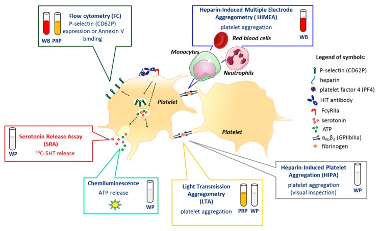
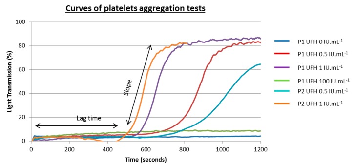
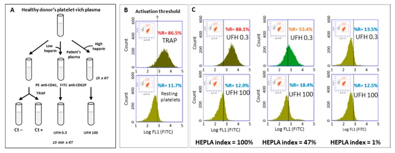
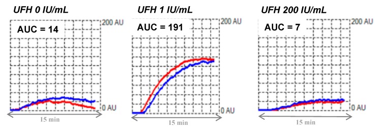
Heparin-induced thrombocytopenia (HIT) is a prothrombotic immune drug reaction caused by platelet-activating antibodies that in most instances recognize platelet factor 4 (PF4)/polyanion complexes. Platelet activation assays (i.e., functional assays) are more specific than immunoassays, since they are able to discern clinically relevant heparin-induced antibodies. All functional assays used for HIT diagnosis share the same principle, as they assess the ability of serum/plasma from suspected HIT patients to activate fresh platelets from healthy donors in the presence of several concentrations of heparin. Depending on the assay, donors' platelets are stimulated either in whole blood (WB), platelet-rich plasma (PRP), or in a buffer medium (washed platelets, WP). In addition, the activation endpoint studied varies from one assay to another: platelet aggregation, membrane expression of markers of platelet activation, release of platelet granules. Tests with WP are more sensitive and serotonin release assay (SRA) is considered to be the current gold standard, but functional assays suffer from certain limitations regarding their sensitivity, specificity, complexity, and/or accessibility. However, the strict adherence to adequate preanalytical conditions, the use of selected platelet donors and the inclusion of positive and negative controls in each run are key points that ensure their performances.

Keywords: diagnosis; functional assays; heparin-induced thrombocytopenia.


Conflict of interest statement

C. Pouplard reports a cooperation contract between Stago and the University of Tours, personal fees from Sobi and Roche, and non-financial support from Sobi, Shire, CSL Behring, Roche, Octapharma, all outside the present work. C. Vayne reports a cooperation contract between Stago and the University of Tours and non-financial support from Shire, Sobi, Roche, CSL Behring, Takeda, all outside the present work. F. Mullier reports institutional fees from Stago, Werfen, Nodia, Sysmex, and Bayer. He also reports speaker fees from Boehringer Ingelheim, Bayer Healthcare, Bristol-Myers Squibb-Pfizer, Stago, Werfen, and Aspen, all outside the present work. T. Lecompte reports a cooperation contract with Stago, outside the present work.

Figures

Figure 1
Figure 1
Platelet responses induced by heparin-induced thrombocytopenia (HIT) antibodies and functional assay targets. Depending on the assay, donors’ platelets are stimulated either in whole blood (WB), platelet-rich plasma (PRP), or after washings (washed platelets, WP). Today, serotonin release assay (SRA), which measures the release of serotonin from dense granules when platelets are activated, and heparin-induced platelet aggregation (HIPA), which is based on a visual inspection of platelet aggregates, are considered the “gold standard” to confirm HIT. The oldest but probably the most used assay is light transmission aggregometry (LTA), classically performed with PRP, but which can also be carried out with WP. Platelet aggregation can also be assessed in whole blood using heparin-induced multiple electrode aggregometry (HIMEA), which considers the possible contribution of monocytes, neutrophils, and red blood cells (RBC) to platelet activation induced by HIT antibodies. Dense granules secretion may also be evaluated by measuring the ATP released using chemiluminescence. Several assays based on flow cytometry (FC) have also been proposed, most of them assessing P-selectin (CD62P) expression or Annexin V binding to phosphatidylserine on the surface of activated platelets.
Figure 2
Figure 2
Typical tracings obtained with light transmission aggregometry in the absence and in the presence of low (0.5 and 1 IU/mL) and high (10 IU/mL) unfractionated heparin UFH concentrations. Aggregation curves for two patients with HIT (P1 and P2) are presented here. Different lag times related to heparin concentrations and patient samples are presented. P1 antibodies appear to be more reactive than P2 antibodies.
Figure 3
Figure 3
Detection of heparin-dependent platelet-activating antibodies with FCA. (A) Schematic procedure of the FCA. (B) The first gate delineates the platelets ((log SSC + log FL2-PE (CD41+ events)) and the second gate is applied to analyze the activated platelets (log FL1-FITC). Determination of the activation threshold: upper panel, TRAP-activated platelets (Ctl+); lower panel, resting platelets (Ctl−), the cursor indicating the activation threshold is placed at the intersection of the FL1 histograms of the positive control (Ctl+) and the negative control (Ctl−). “%R” represents the percentage of CD-62P positive events as an index of platelet activation. This set-up allows determining percentage of the activated platelets (to the right of the activation threshold) under different conditions. (C) Incubations with patients’ plasma and one platelet donor: typical results with low (0.3 IU/mL heparin) and high (100 IU/mL heparin) concentrations of heparin (top and bottom rows, respectively). Left panels: platelets activated with a highly HIT-positive plasma (optical density, OD = 2.5); middle panels: platelets activated with a weakly HIT-positive plasma (OD = 1.3); right panels: platelets incubated with a non-HIT plasma. The HEPLA index is calculated as follows: HEPLA index = (% H 0.3 − % H 100)/(% TRAP Ctl+ − % PBS Ctl−) × 100. FCA: flow cytometric assay, SSC: sideways scatter, PE: phycoerythrin, FITC: fluorescein isothiocyanate, TRAP: thrombin receptor agonist peptide, UFH: unfractionated heparin.
Figure 4
Figure 4
Typical tracing obtained by whole blood impedance aggregometry. Presence of HIT antibodies activating platelets: typical tracings obtained by whole blood impedance aggregometry (Multiplate®) in the absence and in the presence of low (1 IU/mL) and high (200 IU/mL) concentrations of UFH. The whole blood aggregation is recorded for 15 min. AUC: area under curve.

References

    1. Greinacher A., Kohlmann T., Strobel U., Sheppard J.A., Warkentin T.E. The temporal profile of the anti-PF4/heparin immune response. Blood. 2009;113:4970–4976. doi: 10.1182/blood-2008-08-173062. - DOI - PubMed
    1. Cines D.B., Tomaski A., Tannenbaum S. Immune endothelial-cell injury in heparin-associated thrombocytopenia. N. Engl. J. Med. 1987;316:581–589. doi: 10.1056/NEJM198703053161004. - DOI - PubMed
    1. Pouplard C., Iochmann S., Renard B., Herault O., Colombat P., Amiral J., Gruel Y. Induction of monocyte tissue factor expression by antibodies to heparin-platelet factor 4 complexes developed in heparin-induced thrombocytopenia. Blood. 2001;97:3300–3302. doi: 10.1182/blood.V97.10.3300. - DOI - PubMed
    1. Arepally G.M. Heparin-induced thrombocytopenia. Blood. 2017;129:2864–2872. doi: 10.1182/blood-2016-11-709873. - DOI - PMC - PubMed
    1. Perdomo J., Leung H.H.L., Ahmadi Z., Yan F., Chong J.J.H., Passam F.H., Chong B.H. Neutrophil activation and NETosis are the major drivers of thrombosis in heparin-induced thrombocytopenia. Nat. Commun. 2019;10:1322. doi: 10.1038/s41467-019-09160-7. - DOI - PMC - PubMed