. 2020 Apr 24;12(4):391.
doi: 10.3390/pharmaceutics12040391.

HER2-Specific Pseudomonas Exotoxin A PE25 Based Fusions: Influence of Targeting Domain on Target Binding, Toxicity, and In Vivo Biodistribution

Affiliations

HER2-Specific Pseudomonas Exotoxin A PE25 Based Fusions: Influence of Targeting Domain on Target Binding, Toxicity, and In Vivo Biodistribution

Haozhong Ding et al. Pharmaceutics. .

Abstract

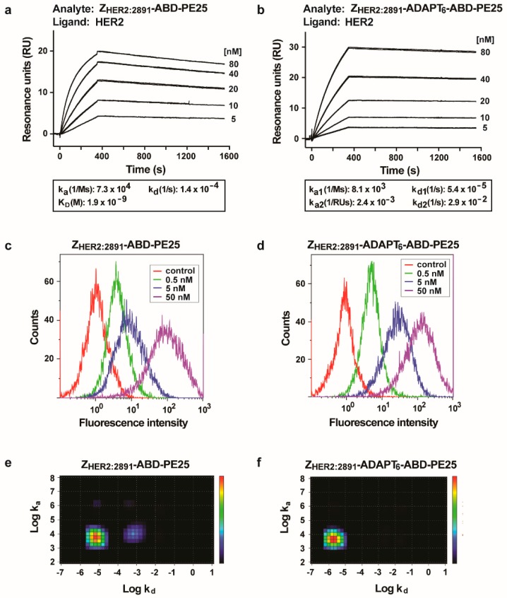
The human epidermal growth factor receptor 2 (HER2) is a clinically validated target for cancer therapy, and targeted therapies are often used in regimens for patients with a high HER2 expression level. Despite the success of current drugs, a number of patients succumb to their disease, which motivates development of novel drugs with other modes of action. We have previously shown that an albumin binding domain-derived affinity protein with specific affinity for HER2, ADAPT6, can be used to deliver the highly cytotoxic protein domain PE25, a derivative of Pseudomonas exotoxin A, to HER2 overexpressing malignant cells, leading to potent and specific cell killing. In this study we expanded the investigation for an optimal targeting domain and constructed two fusion toxins where a HER2-binding affibody molecule, ZHER2:2891, or the dual-HER2-binding hybrid ZHER2:2891-ADAPT6 were used for cancer cell targeting. We found that both targeting domains conferred strong binding to HER2; both to the purified extracellular domain and to the HER2 overexpressing cell line SKOV3. This resulted in fusion toxins with high cytotoxic potency toward cell lines with high expression levels of HER2, with EC50 values between 10 and 100 pM. For extension of the plasma half-life, an albumin binding domain was also included. Intravenous injection of the fusion toxins into mice showed a profound influence of the targeting domain on biodistribution. Compared to previous results, with ADAPT6 as targeting domain, ZHER2:2891 gave rise to further extension of the plasma half-life and also shifted the clearance route of the fusion toxin from the liver to the kidneys. Collectively, the results show that the targeting domain has a major impact on uptake of PE25-based fusion toxins in different organs. The results also show that PE25-based fusion toxins with high affinity to HER2 do not necessarily increase the cytotoxicity beyond a certain point in affinity. In conclusion, ZHER2:2891 has the most favorable characteristics as targeting domain for PE25.

Keywords: HER2; affibody molecule; cancer; half-life extension; pseudomonas exotoxin A.


Conflict of interest statement

The authors declare no conflict of interest.

Figures

Figure 1
Figure 1
Schematic description and initial biochemical characterization of the fusion toxins. (a) A schematic description of the fusion toxins analyzed in the current study along with their theoretical molecular weights. The individual domains of the fusion toxins were connected with linkers with the amino acid sequence (S4G)4. After purification by affinity chromatography with immobilized human serum albumin (HSA), the fusion toxins were analyzed by sodium dodecyl sulfate–polyacrylamide gel electrophoresis (SDS-PAGE) under reducing conditions (b). The numbers to the left indicate molecular weights of reference proteins. The fusion toxins were also analyzed by analytical size exclusion chromatography and the recorded chromatograms are shown in (c). The numbers above the chromatograms correspond to elution volumes of reference proteins.
Figure 2
Figure 2
Analysis of the interaction with HER2. (a,b) Sensorgrams after analyses using a Biacore real-time biosensor of the interaction of the fusion toxins with HER2. Dilution series of the fusion toxins were injected sequentially over a surface with immobilized HER2. Each concentration was injected twice, and each panel is an overlay of both repeats of all concentrations. The injected concentrations are indicated to the right of each panel. SKOV3 cells were stained with different concentrations of the fusion toxins, followed by staining with fluorescently labeled human serum albumin (HSA) and were analyzed by flow cytometry (c,d). The concentration of the fusion toxins used for staining is indicated in the legend. Each panel is an overlay of the plots obtained for all concentrations for each fusion toxin. The controls are non-stained cells. Further, the fusion toxins were radiolabeled with 99mTc and incubated with SKOV3 cells and cell bound radioactivity was recorded as a function of time in a Ligandtracer instrument. The obtained curves were analyzed by Tracedrawer software and the resulting Interaction Maps are presented in (e,f).
Figure 3
Figure 3
Analysis of the interaction between the fusion toxins and HSA and mouse serum albumin (MSA). The analyses were carried out a Biacore real-time biosensor instrument. A dilution series of ZHER2:2891-ABD-PE25 was injected over a surfaces with immobilized HSA (a); a dilution series of ZHER2:2891-ADAPT6-ABD-PE25 was injected over a surface with immobilized HSA (b); a dilution series of ZHER2:2891-ABD-PE25 was injected over a surfaces with immobilized MSA (c); a dilution series of ZHER2:2891-ADAPT6-ABD-PE25 was injected over a surface with immobilized MSA (d). The concentrations are indicated to the right of each panel. Each concentration was injected twice and each panel is an overlay of the sensorgrams recorded for both repeats of each concentration for the whole dilution series.
Figure 4
Figure 4
Determination of cytotoxic potency of fusion toxins. SKBR-3 (a), AU565 (b), SKOV3 (c) and A549 (d) cells were incubated with different concentrations of the fusion toxins and the viability of the cells was measured as a function of protein concentration. The viability of cells incubated without toxin (control cells) was set to 100%. The measured viability was plotted as percent viability compared to control cells on the y-axis, as a function of the concentration of fusion toxin on the x-axis. Each data-point corresponds to the average viability of four independent experiments. The error bars correspond to 1 SD.

References

    1. Emde A., Köstler W.J., Yarden Y. Therapeutic strategies and mechanisms of tumorigenesis of HER2-overexpressing breast cancer. Crit. Rev. Oncol. Hematol. 2012;84:49–57. doi: 10.1016/j.critrevonc.2010.09.002. - DOI - PMC - PubMed
    1. Yan M., Schwaederle M., Arguello D., Millis S.Z., Gatalica Z., Kurzrock R. HER2 expression status in diverse cancers: Review of results from 37,992 patients. Cancer Metastasis Rev. 2015;34:157–164. doi: 10.1007/s10555-015-9552-6. - DOI - PMC - PubMed
    1. Citri A., Yarden Y. EGF-ERBB signalling: Towards the systems level. Nat. Rev. Mol. Cell Biol. 2006;7:505–516. doi: 10.1038/nrm1962. - DOI - PubMed
    1. Slamon D.J., Clark G.M., Wong S.G., Levin W.J., Ullrich A., McGuire W.L. Human breast cancer: Correlation of relapse and survival with amplification of the HER-2/neu oncogene. Science. 1987;235:177–182. doi: 10.1126/science.3798106. - DOI - PubMed
    1. Vogel C.L., Cobleigh M.A., Tripathy D., Gutheil J.C., Harris L.N., Fehrenbacher L., Slamon D.J., Murphy M., Novotny W.F., Burchmore M., et al. Efficacy and Safety of Trastuzumab as a Single Agent in First-Lin Treatment of HER2-Overexpressing Metastatic Breast Cancer. J. Clin. Oncol. 2003;20:719–726. doi: 10.1200/JCO.2002.20.3.719. - DOI - PubMed