. 2020 Apr 28;22(1):95.
doi: 10.1186/s13075-020-2104-7.

Self-reactive T cells induce and perpetuate chronic relapsing arthritis

Affiliations

Self-reactive T cells induce and perpetuate chronic relapsing arthritis

Jonatan Tuncel et al. Arthritis Res Ther. .

Abstract

Background: CD4+ T cells play a central role during the early stages of rheumatoid arthritis (RA), but to which extent they are required for the perpetuation of the disease is still not fully understood. The aim of the current study was to obtain conclusive evidence that T cells drive chronic relapsing arthritis.

Methods: We used the rat pristane-induced arthritis model, which accurately portrays the chronic relapsing-remitting disease course of RA, to examine the contribution of T cells to chronic arthritis.

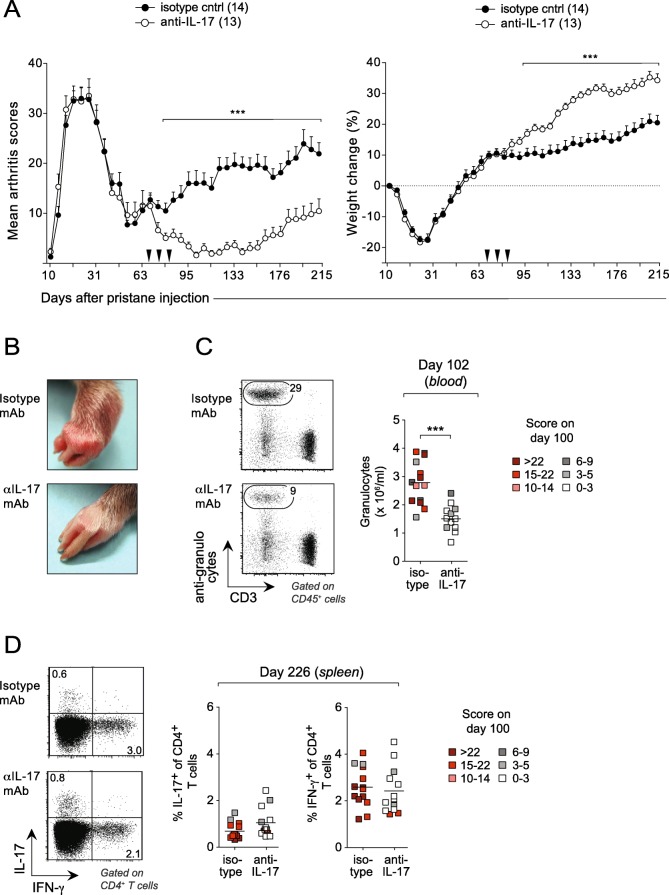
Results: Rats subjected to whole-body irradiation and injected with CD4+ T cells from lymph nodes of pristane-injected donors developed chronic arthritis that lasted for more than 4 months, whereas T cells from the spleen only induced acute disease. Thymectomy in combination with irradiation enhanced the severity of arthritis, suggesting that sustained lymphopenia promotes T cell-driven chronic inflammation in this model. The ability of T cells to induce chronic arthritis correlated with their expression of Th17-associated transcripts, and while depletion of T cells in rats with chronic PIA led to transient, albeit significant, reduction in disease, neutralization of IL-17 resulted in almost complete and sustained remission.

Conclusion: These findings show that, once activated, self-reactive T cells can sustain inflammatory responses for extended periods of time and suggest that such responses are promoted in the presence of IL-17.

Keywords: Adoptive T cell transfer; Chronic arthritis; MHC class II; PIA; Pristane; RA; T cell depletion.


Conflict of interest statement

PO, MH, and RH have financial interest in the company Redoxis AB that provides research service in the area of preclinical models including the model described in this article. The other authors declare that they have no competing interests.

Figures

Fig. 1
Fig. 1
Joint-draining lymph node-derived CD4+ T cells from pristane-injected rats induce chronic arthritis. a Arthritis development in irradiated rats transferred with 2 × 10⁷ in vitro-re-stimulated cells from the LN or spleen of pristane-injected donors (14 days after pristane injection). Control rats were transferred with an equal number of non-re-stimulated LN cells. n = 4 rats/group. b Chronic relapses of arthritis in individual paws of a representative recipient transferred with re-stimulated LN cells. n = 1. c H&E staining of a representative arthritic hind paw (top) showing typical pannus formation above the joint cavity at day 124 after injection of re-stimulated cells from LNs of pristane-injected donors. Bottom image shows a corresponding section from a rat transferred with non-re-stimulated cells. d Serum levels of COMP and AGP on day 124 post-transfer. Control, rats transferred with non-re-stimulated LN cells; PIA, rats with chronic PIA (non-transferred). n = 4–6/group. e Arthritis development in irradiated rats transferred with 2 × 10⁷ in vitro-re-stimulated CD4+ T cells. n = 3. Data show mean values ± SD. Statistical analyses using the Mann-Whitney U test; *< 0.05, **< 0.01. RH, right hind paw; LH, left hind paw; RF, right front paw; LF, left front paw
Fig. 2
Fig. 2
Irradiation in combination with thymectomy enhances chronic arthritis. a Rats were pre-conditioned by irradiation and/or thymectomy (as indicated in the enclosed table) prior to transfer. Left: arthritis development in rats transferred with or without LN-derived cells. Right: summary data of total incidence (Incid), incidence of arthritis lasting > 40 days (Chronic), and level of acute-phase protein (AGP). Number of rats per group is indicated in the enclosed table. b Immunohistochemistry of hind paws from rats shown in a stained for CD4+ cells, macrophages and neutrophils. Bottom tables show quantifications (as percent of total cells) of infiltrating cell populations; 0.5 to 5% (++), 5 to 20% (+++), or 20 to 50% (++++). c Irradiated/thymectomized rats (n = 70) were transferred with T cells as in a. On day 65 post-transfer, rats were divided into three disease-matched groups that were treated with methotrexate (n = 9), etanercept (n = 9), or phytol (n = 8). Filled and open circles represent normalized disease scores (vs. day 65) for treated and none-treated individuals, respectively. Statistical analyses as in Fig. 1. All groups in a were compared to non-irradiated, non-thymectomized rats except those indicated by a hash sign (#), which were compared to irradiated, non-thymectomized recipients. Data shown in c were pooled from two separate experiments
Fig. 3
Fig. 3
The ability of CD4+ T cells to transfer chronic arthritis correlates with their expression of Th17-associated cytokines. a Top: representative flow cytometric plots of CD4+ T cells from various lymphoid organs of pristane-injected rats (14 days after pristane injection) before and after in vitro isolation. Bottom: transcript levels, determined by quantitative RT-PCR and depicted as fold change (FC), of various cytokines in CD4+ T cells from lymph node and spleen, before (–) and 14 days after (+) injection of pristane. The expression of each target gene was normalized to the geometric mean of the expression of Arbp, Gusb, and Actb. n = 4 rats/group. b Arthritis development in rats transferred with 2 × 10⁷ in vitro-re-stimulated cells from inguinal or mesenteric LNs (n = 5–9 rats/group) of pristane-injected donors. c Corresponding data (as in a) for various transcription factors. Box and whisker plots in a show upper and lower quartiles (the outer boundaries of the box), median (horizontal line inside box) and highest and lowest observations (whiskers). Data in c shows fold change ± SD. Statistical analyses using the Mann-Whitney U test; *< 0.05, **< 0.01.1, ***< 0.001. iLN, inguinal lymph nodes; mLN, mesenteric lymph nodes; Spl, spleen
Fig. 4
Fig. 4
T cell ablation in rats with chronic PIA temporarily ameliorates the progression of disease. a Rats with early-stage chronic arthritis were treated with a mAb to αβTCR, or an isotype-matched control mAb, on day 67, 76, and 82 after pristane injection (arrow heads). Left: clinical scores. Right: relative weight change as compared to day 0. n = 13–14 rats/group. b Left: representative flow cytometric plots of peripheral blood mononuclear cells with gates depicting frequency of CD3+ cells (left) and CD4+ T cells (right) in T cell-depleted (bottom) and non-depleted (upper) rats on day 102 after pristane injection. Right: summary data of CD3+ and CD4+ T cell titers. The shading of the symbols represents the severity of arthritis on day 100 after pristane injection according to the enclosed legend. Data in a show mean values ± SD. Horizontal line in b depicts mean values. Statistical analyses as in Fig. 1
Fig. 5
Fig. 5
Neutralization of IL-17 efficiently blocks the progression of chronic disease in PIA. a Rats, which were part of the same cohort as those shown in Fig. 4, were treated with a mAb to IL-17, or an isotype-matched control mAb, on day 67, 76, and 82 after pristane injection (arrow heads). Left: clinical scores. Right: relative weight change as compared to day 0. n = 13–14 rats/group. b Representative images of front paws of rats injected with pristane (day 102 after injection) and treated with either IL-17 depleting or isotype control antibodies. c Left: representative flow cytometric plots of peripheral blood mononuclear cells with gates depicting frequency of granulocytes in IL-17-depleted (bottom) and non-depleted (upper) rats on day 102 after pristane injection. Right: summary data of granulocyte titers. d Representative flow cytometric plots and summary data for the percentage of IL-17+ and IFNγ+ CD4+ T cells on day 226 in the spleen. The colors of the symbols in c and d represent the severity of arthritis on day 100 after pristane injection according to the enclosed legends. Data in a show mean values ± SD. Horizontal line in c and d depicts mean values. Statistical analyses as in Fig. 1

References

    1. Kim K, Bang S-Y, Lee H-S, Cho S-K, Choi C-B, Sung Y-K, et al. High-density genotyping of immune loci in Koreans and Europeans identifies eight new rheumatoid arthritis risk loci. Ann Rheum Dis. 2015;74:e13. doi: 10.1136/annrheumdis-2013-204749. - DOI - PMC - PubMed
    1. Okada Y, Terao C, Ikari K, Kochi Y, Ohmura K, Suzuki A, et al. Meta-analysis identifies nine new loci associated with rheumatoid arthritis in the Japanese population. Nat Genet. 2012;44:511–516. doi: 10.1038/ng.2231. - DOI - PubMed
    1. Stahl EA, Raychaudhuri S, Remmers EF, Xie G, Eyre S, Thomson BP, et al. Genome-wide association study meta-analysis identifies seven new rheumatoid arthritis risk loci. Nat Genet. 2010;42:508–514. doi: 10.1038/ng.582. - DOI - PMC - PubMed
    1. Plenge RM, Padyukov L, Remmers EF, Purcell S, Lee AT, Karlson EW, et al. Replication of putative candidate-gene associations with rheumatoid arthritis in >4,000 samples from North America and Sweden: association of susceptibility with PTPN22, CTLA4, and PADI4. Am J Hum Genet. 2005;77:1044–1060. doi: 10.1086/498651. - DOI - PMC - PubMed
    1. The RACI consortium, the GARNET consortium, Okada Y, Wu D, Trynka G, Raj T, et al. Genetics of rheumatoid arthritis contributes to biology and drug discovery. Nature. 2013;506:376–381. - PMC - PubMed

Publication types