. 2020 Jun 5;295(23):7970-7980.
doi: 10.1074/jbc.RA120.012658. Epub 2020 Apr 28.

The F-box protein FBXL16 up-regulates the stability of C-MYC oncoprotein by antagonizing the activity of the F-box protein FBW7

Affiliations

The F-box protein FBXL16 up-regulates the stability of C-MYC oncoprotein by antagonizing the activity of the F-box protein FBW7

Marion Morel et al. J Biol Chem. .

Abstract

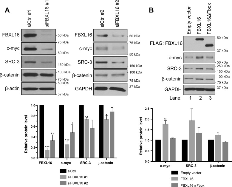
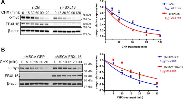
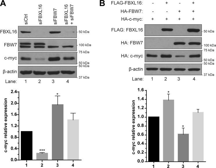
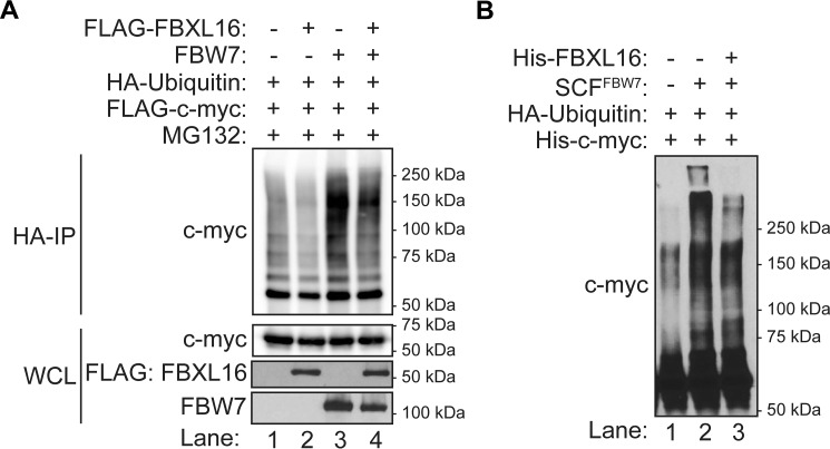
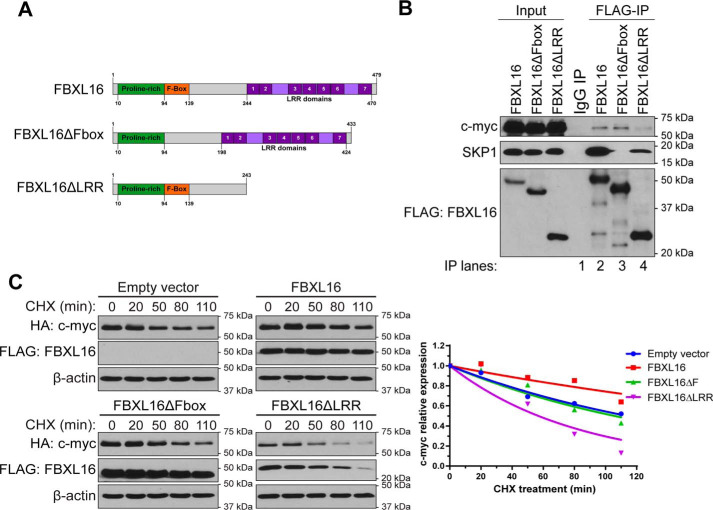
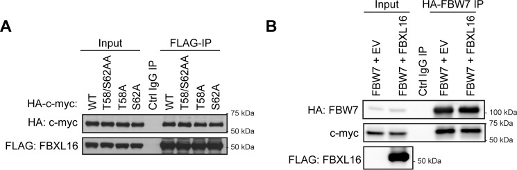
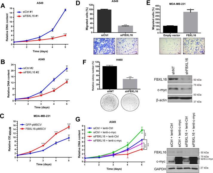
F-box proteins, such as F-box/WD repeat-containing protein 7 (FBW7), are essential components of the SKP1-CUL1-F-box (SCF) E3 ubiquitin ligases. They bind to S-phase kinase-associated protein 1 (SKP1) through the F-box motif and deliver their protein substrate to the E3 ligase complex for ubiquitination and subsequent degradation. F-box and leucine-rich repeat protein 16 (FBXL16) is a poorly studied F-box protein. Because it does not interact with the scaffold protein cullin 1 (CUL1), we hypothesized that FBXL16 might not form a functional SCF-E3 ligase complex. In the present study, we found that FBXL16 up-regulates the levels of proteins targeted by SCF-E3 ligases, such as C-MYC, β-catenin, and steroid receptor coactivator 3 (SRC-3). Focusing on C-MYC, a well-known oncoprotein overexpressed in most human cancers, we show that FBXL16 stabilizes C-MYC by antagonizing FBW7-mediated C-MYC ubiquitination and degradation. Further, we found that, although FBXL16 does not interact with CUL1, it interacts with SKP1 via its N-terminal F-box domain and with its substrate C-MYC via its C-terminal leucine-rich repeats (LRRs) domain. We found that both the F-box domain and the LRR domain are important for FBXL16-mediated C-MYC stabilization. In line with its role in up-regulating the levels of the C-MYC and SRC-3 oncoproteins, FBXL16 promoted cancer cell growth and migration and colony formation in soft agar. Our findings reveal that FBXL16 is an F-box protein that antagonizes the activity of another F-box protein, FBW7, and thereby increases C-MYC stability, resulting in increased cancer cell growth and invasiveness.

Keywords: E3 ubiquitin ligase; F-box and leucine-rich repeat protein 16 (FBXL16); F-box protein; F-box/WD repeat-containing protein 7 (FBW7); Myc (c-Myc); cancer; cell migration; protein homeostasis; protein stability; proto-oncogene C-Myc (C-MYC); ubiquitylation (ubiquitination).


Conflict of interest statement

Conflict of interest—The authors declare that they have no conflicts of interest with the contents of this article.

Figures

Figure 1.
Figure 1.
FBXL16 up-regulates the protein levels of SCF E3 ligase substrates. A, A549 cells were transiently transfected with a negative control siRNA (siCtrl #1 or siCtrl #2) or an siRNA targeting FBXL16 (siFBXL16 #1 or siFBXL16 #2). Western blot analysis and quantification shows a decrease in c-myc, SRC-3, and β-catenin protein levels upon FBXL16 knockdown. B, Western blotting analyses of FLAG-FBXL16 (or FLAG-FBXL16ΔFbox) using an anti-FLAG Ab, c-myc, SRC-3, β-catenin, and β-actin in H1299 cells transiently transfected with a control empty vector, a vector expressing FLAG-tagged FBXL16 protein, or a vector expressing FLAG-tagged FBXL16 mutant with the deletion of F-box domain (FBXL16ΔFbox). H1299 cells were treated with CHX for 15 min prior to being lysed. Western blots are representative of three independent experiments. Values in graph bars represent mean ± S.E. and statistical significances were determined by Student's t test (*, p < 0.05; **, p < 0.01; ***, p < 0.001).
Figure 2.
Figure 2.
FBXL16 increases c-myc protein stability. A, A549 cells were transiently transfected with a negative control siRNA (siCtrl) or an siRNA targeting FBXL16 (siFBXL16). 48 h post transfection, protein translation was inhibited with CHX (100 μg/ml) for different times as indicated, followed by cell lysis and Western blot analysis of protein levels. c-myc protein level at each time point was normalized to that of β-actin, and the normalized c-myc protein level at 0 min time point was arbitrarily set as 1. c-myc half-life (t½) was determined from the exponential curve equation calculated using the one-phase exponential decay model (GraphPad Prism 6 software). B, MDA-MB-231 stable cells overexpressing pMSCV-GFP or pMSCV-FBXL16 were treated with CHX for different times and the half-life of endogenous c-myc protein was determined as in (A).
Figure 3.
Figure 3.
FBXL16 regulates c-myc by antagonizing FBW7. A, Western blot analysis and quantification of c-myc protein level in A549 cells transiently transfected with negative control siRNAs (siCtrl), siRNA targeting FBW7 (siFBW7), siRNA targeting FBXL16 (siFBXL16), or both siFBW7 and siFBXL16. B, 293T cells were transiently co-transfected with HA–c-myc together with either empty plasmid controls, FLAG-FBXL16, HA-FBW7, or both FLAG-FBXL16 and HA-FBW7, followed by Western blot analysis using an anti-FLAG Ab for measuring FBXL16 and an anti-HA Ab for measuring both c-myc and FBW7. Western blots are representative of three independent experiments. Values in graph bars represent mean ± S.E. and statistical significances were determined by Student's t test (*, p < 0.05; **, p < 0.01; ***, p < 0.001).
Figure 4.
Figure 4.
FBXL16 decreases FBW7-depandant c-myc ubiquitination. A, 293T cells were co-transfected with pMT-HA-ubiquitin and pCMV-FLAG-c-myc plasmids together with either pSG5-FLAG-FBXL16, pcDNA-FBW7, or both as indicated. HA-ubiquitin conjugates were immunoprecipitated using anti-HA affinity gel and then immunoblotted with anti–c-myc antibody to detect the ubiquitinated c-myc proteins. The expression levels of c-myc, FBXL16, and FBW7 in whole cell lysate (WCL) were immunoblotted with anti–c-myc, anti-FLAG, and anti-FBW7 antibodies, respectively. B, in vitro ubiquitination assay was performed by incubating the purified recombinant c-myc protein with HA-ubiquitin, E1 and E2 enzymes together with or without SCFFBW7, and/or FBXL16 for 1 h at 37 °C, followed by Western blot analysis using anti–c-myc antibody.
Figure 5.
Figure 5.
Both F-box and LRR domains of FBXL16 are important for the regulation of c-myc. A, schematic structures of full-length FBXL16 protein, FBXL16 with the deletion of F-box domain (FBXL16ΔFbox), and FBXL16 with the deletion of the c-terminal LRRs (FBXL16ΔLRR). The numbers below or above the structures indicate the positions of the amino acids. B, FLAG-tagged FBXL16, FBXL16ΔFbox, or FBXL16ΔLRR was overexpressed in 293T cells, followed by immunoprecipitation using anti–FLAG-Ab conjugated agarose beads (FLAG-IP). Western blot analysis was then performed to examine the interactions of FBXL16 or the deletion mutant with endogenous SKP1 and c-myc proteins. C, HeLa cells were transiently co-transfected with HA–c-myc together with either an empty vector control, FLAG-FBXL16, FLAG-FBXL16ΔFbox, or FLAG-FBXL16ΔLRR. 32 h post transfection, protein translation was inhibited with CHX (100 μg/ml) for different times (minutes), followed by cell lysis and Western blot analysis of protein levels. c-myc protein level at each time point was normalized to that of β-actin, and the normalized c-myc protein level at 0 min time point was arbitrarily set as 1. Exponential curves were extrapolated using the one-phase exponential decay model (GraphPad Prism 6 software).
Figure 6.
Figure 6.
FBXL16 does not compete with FBW7 for c-myc binding. A, 293T cells were co-transfected with pSG5-FLAG-FBXL16 and either WT, T58/S62AA, T58A, or S62A of HA-tagged c-myc constructs. FLAG-FBXL16 was immunoprecipitated using anti–FLAG-Ab conjugated agarose beads (FLAG-IP), and then Western blot analysis was performed to examine the interactions of FBXL16 with different HA-tagged c-myc protein. B, HA-tagged FBW7 was co-overexpressed with either an empty plasmid or pSG5-FLAG-FBXL16 in 293T cells. HA-FBW7 was then immunoprecipitated using anti–HA-Ab conjugated agarose beads (HA-FBW7 IP). Western blot analysis was then performed to examine the interactions of FBW7 with c-myc.
Figure 7.
Figure 7.
FBXL16 promotes cancer cell growth and migration. A, cell growth was determined by dsDNA quantification. After transient transfection of A549 cells with a negative control siRNA (siCtrl #1) or an siRNA targeting FBXL16 (siFBXL16 #1), DNA content was measured every 24 h and values were normalized to day 1. B, A549 cells were transiently transfected with siCtrl#2 or siFBXL16#2 and cell growth was assessed as in (A). C, an MTS proliferation assay was performed for MDA-MB-231 stable cells overexpressing pMSCV-GFP or pMSCV-FBXL16. Cell viability was determined by measuring A490 nm every 24 h and values were normalized to day 1. A–C, values in graphs represent mean ± S.D. of a representative experiment and statistical significances were determined by two-way ANOVA (*, p < 0.05; **, p < 0.01; ***, p < 0.001). D, A549 cells were transfected with siCtrl or siFBXL16 and their ability to migrate was analyzed by a two-chamber Transwell assay. Migrated cells were stained and photographed for quantification by counting. The migration ability of cells treated with siFBXL16 is presented as the percentage of the number of migrated cells per field in siFBXL16 over that of siCtrl. E, MDA-MB-231 cells were transiently transfected with an empty vector control or FBXL16 and their ability to migrate was analyzed as in (D). D and E, values in the bar graphs represent mean ± S.E. of three independent experiments. Statistical significance was determined by Student's t test (*, p < 0.05; **, p < 0.01; ***, p < 0.001). Representative images are shown at the bottom of the graphs. Scale bars, 100 μm. F, stable H460 cell lines with inducible expression of a nontargeting control shRNA (shNT) or a shRNA targeting FBXL16 (shFBXL16) were grown in soft agar in presence of doxycycline (0.5 μg/ml). After 8 days, colonies were stained and quantified using ImageJ software. The colony formation relative to shNT was determined from the average of two experiments with three replicates each time. Values in bar graphs represent mean ± S.E. Representative wells are shown at the bottom of the graphs. On the right, FBXL16 knockdown was confirmed by Western blot analysis after 5 days of doxycycline treatment (0.5 μg/ml). G, A549 cells were transiently transfected with siCtrl or siFBXL16 and transduced with a control lentivirus (lenti-Ctrl) or lentiviral FLAG-tagged c-myc (lenti-c-myc). Cell growth was assessed by dsDNA quantification as in (A). Values in graph represent mean ± S.D. of a representative experiment. Statistical significance on day 5 was determined by two-way ANOVA (*, p < 0.05; **, p < 0.01; ***, p < 0.001). On the right, knockdown of FBXL16 and lentiviral overexpression of FLAG-tagged c-myc (lenti-c-myc) were confirmed by Western blot analysis 2 days post transfection.

References

    1. Hershko A., and Ciechanover A. (1998) The ubiquitin system. Annu. Rev. Biochem. 67, 425–479 10.1146/annurev.biochem.67.1.425 - DOI - PubMed
    1. Pickart C. M. (2001) Mechanisms underlying ubiquitination. Annu. Rev. Biochem. 70, 503–533 10.1146/annurev.biochem.70.1.503 - DOI - PubMed
    1. Dye B. T., and Schulman B. A. (2007) Structural mechanisms underlying posttranslational modification by ubiquitin-like proteins. Annu. Rev. Biophys. Biomol. Struct. 36, 131–150 10.1146/annurev.biophys.36.040306.132820 - DOI - PubMed
    1. Zheng N., and Shabek N. (2017) Ubiquitin ligases: Structure, function, and regulation. Annu. Rev. Biochem. 86, 129–157 10.1146/annurev-biochem-060815-014922 - DOI - PubMed
    1. Petroski M. D., and Deshaies R. J. (2005) Function and regulation of cullin-RING ubiquitin ligases. Nat. Rev. Mol. Cell Biol. 6, 9–20 10.1038/nrm1547 - DOI - PubMed

Publication types