. 2021 Jan;70(1):55-66.
doi: 10.1136/gutjnl-2019-319748. Epub 2020 Apr 27.

YAP1 mediates gastric adenocarcinoma peritoneal metastases that are attenuated by YAP1 inhibition

Affiliations

YAP1 mediates gastric adenocarcinoma peritoneal metastases that are attenuated by YAP1 inhibition

Jaffer A Ajani et al. Gut. 2021 Jan.

Erratum in

Abstract

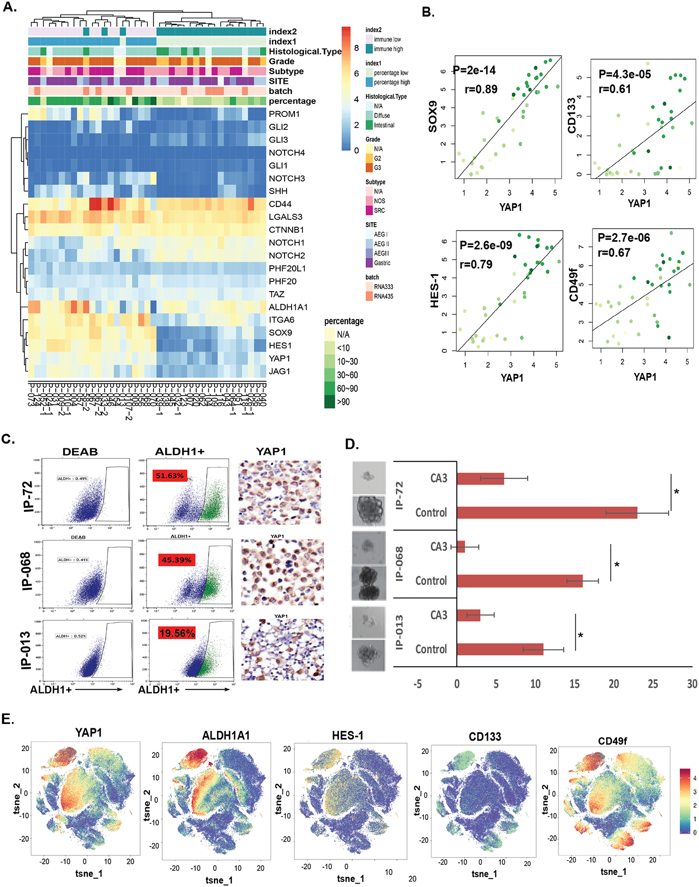
Objective: Peritoneal carcinomatosis (PC; malignant ascites or implants) occurs in approximately 45% of advanced gastric adenocarcinoma (GAC) patients and associated with a poor survival. The molecular events leading to PC are unknown. The yes-associated protein 1 (YAP1) oncogene has emerged in many tumour types, but its clinical significance in PC is unclear. Here, we investigated the role of YAP1 in PC and its potential as a therapeutic target.

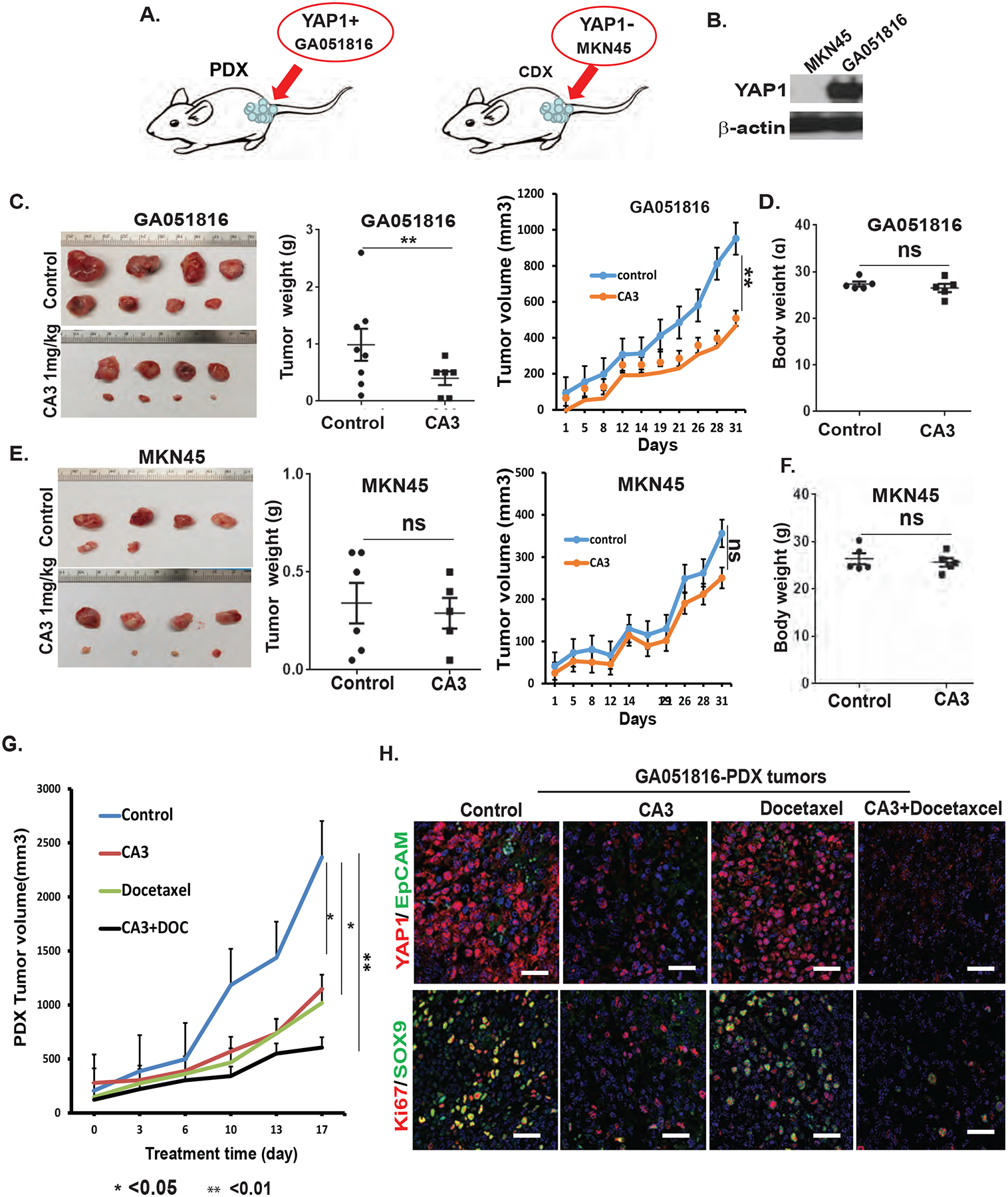
Methods: Patient-derived PC cells, patient-derived xenograft (PDX) and patient-derived orthotopic (PDO) models were used to study the function of YAP1 in vitro and in vivo. Immunofluorescence and immunohistochemical staining, RNA sequencing (RNA-Seq) and single-cell RNA-Seq (sc-RNA-Seq) were used to elucidate the expression of YAP1 and PC cell heterogeneity. LentiCRISPR/Cas9 knockout of YAP1 and a YAP1 inhibitor were used to dissect its role in PC metastases.

Results: YAP1 was highly upregulated in PC tumour cells, conferred cancer stem cell (CSC) properties and appeared to be a metastatic driver. Dual staining of YAP1/EpCAM and sc-RNA-Seq revealed that PC tumour cells were highly heterogeneous, YAP1high PC cells had CSC-like properties and easily formed PDX/PDO tumours but also formed PC in mice, while genetic knockout YAP1 significantly slowed tumour growth and eliminated PC in PDO model. Additionally, pharmacologic inhibition of YAP1 specifically reduced CSC-like properties and suppressed tumour growth in YAP1high PC cells especially in combination with cytotoxics in vivo PDX model.

Conclusions: YAP1 is essential for PC that is attenuated by YAP1 inhibition. Our data provide a strong rationale to target YAP1 in clinic for GAC patients with PC.

Keywords: gastric adenocarcinoma; gene regulation; molecular oncology.


Conflict of interest statement

Competing interests: None declared.

Figures

Figure 1.
Figure 1.. YAP1 is highly expressed only in PC malignant cells and peritoneal biopsy cancer cells
A. CyTOF analysis of PC cell composition in 20 PC samples using epithelial markers EpCAM, cytokeratin and nonepithelial marker-vimentin, CD3 as well as YAP1 as described in Materials Methods. B. Expression of YAP1 was determined by IHC staining using anti-human YAP1 antibody in PC cell block slides (top) and peritoneal biopsy tissue formalin- fixed paraffin-embedded slides (bottom). scale bar:20μm; C. Representative PC cases were stained by dual-immunofluorescent staining of YAP1 and EpCAM, an epithelial tumor marker as described as Materials &Methods; Scale: 2520μm; D. Representative PC cases were co-stained YAP1 with immune marker CD45, macrophage CD163 and stromal marker vimentin by dual-immunofluorescence staining using their antibodies respectively. Scale: 2520μm; E. The association of YAP1 expression with GC patients’ survival in 631 advanced GAC patients from TCGA database (kmplot.com). p<0.001.
Figure 2.
Figure 2.. Heterogeneity in PC cells by YAP1/EpCAM co-staining and sc-RNA Seq.
A. Expression of YAP1 (red) and EpCAM (green) was determined by dual-immunofluorescence staining using anti-mouse EpCAM and anti-YAP1 rabbit antibody in 123 cases of PC. Discrepant expression of YAP1 and EpCAM within and between PC samples was noted. Nuclear YAP1 (red arrow) and membranous EpCAM (green arrow) within a specimen or among PC specimen were demonstrated. B. The numbers of PC cases expressing both YAP1 and EpCAM; and those with high YAP1 or high EpCAM, and expressing only YAP1 or only EpCAM are shown in the table. C. Expression analysis of EpCAM and YAP1 was performed by sc-RNA-Seq in tumor cell clusters from 20 PC samples as described in online Materials&Methods.
Figure 3.
Figure 3.. YAP1 expression is strongly associated with CSC properties in PC specimen by RNA-Seq.
A. RNA-Seq profiling of 39 PC samples using a panel of notable CSC-related genes. Unsupervised hierarchical clustering was performed on the normalized RNA expression data of these genes. B. Significant associations between YAP1 and notable CSC-related markers SOX9, HES1, CD133, and CD49f are depicted in scatter graphs from RNA-Seq data in the 39 PC samples. P values were calculated by the non-parametric Mann-Whitney U test. C. Isolated ascites cells from three PC samples (IP-013, IP-068 and IP-72) were labeled for the ALDH1+ cell population using an ALDH1 labeling kit. Shown are the ALDH1+ percentage of the population in each PC sample and IHC staining of YAP1 in relative PC cell blocks. D. Tumor sphere capacity of these three PC samples treated with or without YAP1 inhibitor CA3 at 0.5μM were determined as described in Materials and Methods. E. Expression of YAP1 and CSC markers ALDH1, HES1, CD133, and CD49f was analyzed in CyTOF data of 10 separated PC samples.
Figure 4.
Figure 4.. YAP1high PC cells easily form PDXs in mice irrespective of PC tumor cell purity or EpCAM expression.
A. Samples harvested from malignant ascites in representative PC cases (IP-116 and IP-013) underwent immunofluorescence (IF) staining of YAP1 and EpCAM and were also injected into nude mice for generation of PDX tumors, which were then stained by hematoxylin and eosin (HE) to determine morphology and subjected to dual-immunofluorescence staining with anti-mouse EpCAM and anti-YAP1 rabbit antibody. B. Successful knockout of YAP1 in GA051816 cells using LentiCRISPR/Cas9. C. Tumor sphere formation (left) and invasion capacity (right) in GA051816 parental cells were compared with that of the YAP1 knockout cells. **p<0.01. Experiments repeat at least three times. D. GA051816 PC cells with or without YAP1 knockout were labeled for ALDH1+ cell population using an ALDH1 labeling kit. The ALDH1+ population in each group is shown in the bar graph. E. Tumorigenicity at serial dilution was assessed in GA051816 control cells compared with YAP1 knockout cells on day 21 after cells inoculation. 10 mice per group. F. Bulk RNA-Seq was performed in GA051816 control cells and YAP1 knockout cells, and critical mediators in CSCs (red arrow) and EMT signaling (green arrow) were identified and depicted.
Figure 5.
Figure 5.. YAP1 is essential for PC metastases in PDO model.
A. GA051816 PC cells transfected with mCherry-Luciferase were visualized under microscope (top). High luciferase activity in mCherry-Luciferase transfected GA051816 cells (bottom)) was confirmed by luciferin. B. mCherry-Luciferase GA051816 control or YAP1 knockout cells were injected into the stomach wall of NOD/SCID mice, and tumor growth in the stomach wall and metastases to the peritoneal cavity or other organs were monitored by bioluminescence weekly. C. Representative image of peritoneal metastasis of GA051816 parental cells and no metastasis in GA051816 YAP1 knockout cells. D. Survival of mice implanted with GA051816 parental cells compared with mice implanted with GA051816 YAP1 knockout cells. E. Analysis of ALDH1 labeling using ALDH1 labeling kit in cells from primary tumor and PC from same mouse. Results are repeated in at least three mice. F. Expression of YAP1, SOX9, and other CSC markers was detected in primary and PC cells from the same mouse using Western blot and quantitative polymerase chain reaction. Tumor sphere formation capacity is depicted as well. G. Association of expression of YAP1 and ALDH1 was detected using dual immunofluorescence in GA051816 PC cells.
Figure 6.
Figure 6.. Pharmacologic inhibition of YAP1 specifically suppress patient-derived cells growth and colony formation in YAP1 high cells
A, Expression of YAP1 was determined by immunoblotting in GA051816 cells treated with CA3 at indicated dosage for 48 hours. B. Tumor sphere formation in GA051816 cells treated with CA3 at different dosages was assessed. C. GA051816 PC cells treated with CA3 at 0.5 μM for 48 hours and then labeled for ALDH1+ cell population using an ALDH1 labeling kit. The ALDH1+ population in each group is shown in the bar graph. Data represent as mean and standard deviation from three experiments. D. Expression of YAP1 was determined by immunoblotting in three additional PC cells from patients (GA070716, GA011017, and GA011317) treated with CA3 at 1 μM for 48 hours (upper). Tumor sphere formation for GA070716, GA011017, and GA011317 cells treated with CA3 at 1 μM for 10 days (lower panel). E. Cell viability in GA051816 parental and YAP1 knockout cells treated with CA3 at the indicated dosage for 3 days. YAP1 high parental cells are more sensitive to CA3. **p<0.01. F&G. Colony formation and tumor sphere formation were assessed in GA051816 (YAP1 high) and MKN45 (YAP1 none) GC cells treated with CA3 at the dosage indicated. Colony number and tumor sphere number were calculated after 14 days. **p< 0.01; ***p<0.0001.
Figure 7.
Figure 7.. Pharmacologic inhibition of YAP1 significantly inhibit PDX tumor growth in YAP1 high PC cells in Vivo.
A. Diagram demonstrates the PDX tumor formation in GA051816 YAP1high and CDX tumor formation in MKN45 GC cell line with no YAP1expression. B. Expression of YAP1 was detected by western blotting in both MNK45 and GA051816 cells; C. Representative tumors in GA051816 PDX tumors and treated with CA3 at 1mg/kg, three time a week for three weeks are shown, and tumor weights, tumor volumes from each group were calculated. D. mouse body weights in GA051816 PDX tumors were calculated (E) Representative tumor images, tumor weight, tumor volume in MKN45 CDX model treated with CA3, three times a week, 1mg/kg, IP injection. F. mouse body weights in MKN45 CDX tumors were calculated. G. YAP1 inhibitor CA3 enhances the effects of docetaxel in inhibiting growth of PDX tumors. GA051816 patient-derived cells (2×10⁶ cells) were injected subcutaneously in nude mice, at two sites (left and right) per mouse and treated with CA3 alone, docetaxel alone, or the combination with five mice per group for three weeks. Tumor volumes were measured twice per week for three weeks and calculated as described in Materials. *P<0.05. **P<0.01. H. Dual immunofluorescence staining of YAP1/EpCAM and Ki67/SOX9 were performed in PDX tumors of four groups as described in Materials and Methods. Scale bar, 20μm.

References

    1. Siegel RL, Miller KD, Jemal A, Cancer statistics, 2019. CA Cancer J Clin 69, 7–34 (2019). - PubMed
    1. Lamar JM, Stern P, Liu H, Schindler JW, Jiang ZG, Hynes RO, The Hippo pathway target, YAP, promotes metastasis through its TEAD-interaction domain. Proc Natl Acad Sci U S A 109, E2441–2450 (2012). - PMC - PubMed
    1. Kitayama J, Intraperitoneal chemotherapy against peritoneal carcinomatosis: current status and future perspective. Surg Oncol 23, 99–106 (2014). - PubMed
    1. Gill RS, Al-Adra DP, Nagendran J, Campbell S, Shi X, Haase E, Schiller D, Treatment of gastric cancer with peritoneal carcinomatosis by cytoreductive surgery and HIPEC: a systematic review of survival, mortality, and morbidity. Journal of surgical oncology 104, 692–698 (2011). - PubMed
    1. Sadeghi B, Arvieux C, Glehen O, Beaujard AC, Rivoire M, Baulieux J, Fontaumard E, Brachet A, Caillot JL, Faure JL, Porcheron J, Peix JL, François Y, Vignal J, Gilly FN, Peritoneal carcinomatosis from non-gynecologic malignancies: results of the EVOCAPE 1 multicentric prospective study. Cancer 88, 358–363 (2000). - PubMed

Publication types

Substances