. 2020 Jul 3;369(6499):eabb9601.
doi: 10.1126/science.abb9601. Epub 2020 Apr 28.

Feasibility of blood testing combined with PET-CT to screen for cancer and guide intervention

Affiliations

Feasibility of blood testing combined with PET-CT to screen for cancer and guide intervention

Anne Marie Lennon et al. Science. .

Abstract

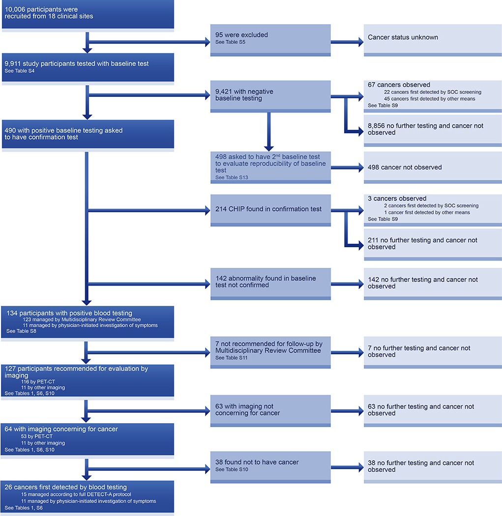
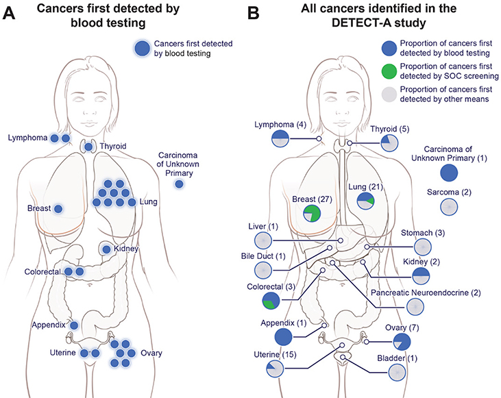
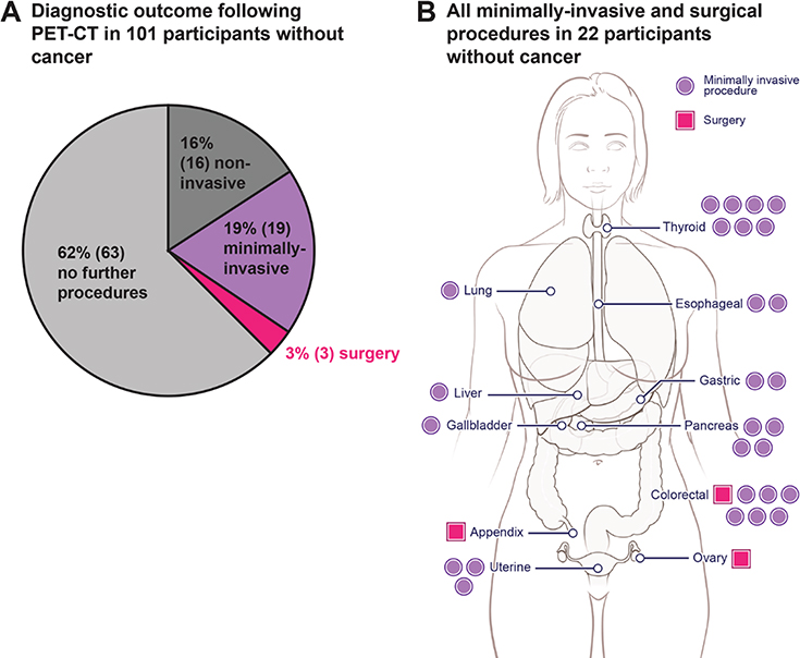
Cancer treatments are often more successful when the disease is detected early. We evaluated the feasibility and safety of multicancer blood testing coupled with positron emission tomography-computed tomography (PET-CT) imaging to detect cancer in a prospective, interventional study of 10,006 women not previously known to have cancer. Positive blood tests were independently confirmed by a diagnostic PET-CT, which also localized the cancer. Twenty-six cancers were detected by blood testing. Of these, 15 underwent PET-CT imaging and nine (60%) were surgically excised. Twenty-four additional cancers were detected by standard-of-care screening and 46 by neither approach. One percent of participants underwent PET-CT imaging based on false-positive blood tests, and 0.22% underwent a futile invasive diagnostic procedure. These data demonstrate that multicancer blood testing combined with PET-CT can be safely incorporated into routine clinical care, in some cases leading to surgery with intent to cure.


Conflict of interest statement

Competing interests: B.V., K.W.K., N.P., I.K., and C.L. are founders of and hold equity in Thrive Earlier Detection. K.W.K., N.P., and C.L. are consultants to and are on the Board of Directors of, Thrive Earlier Detection. C.L. is an officer of Thrive Earlier Detection. B.V., K.W.K., N.P. & S.Z. is a founder of, holds equity in, and serves as a consultant to Personal Genome Diagnostics. S.Z. holds equity in Thrive Earlier Detection, and has a research agreement with BioMed Valley Discoveries, Inc. K.W.K. & B.V. are consultants to Sysmex, Eisai, and CAGE Pharma and hold equity in CAGE Pharma. B.V. is also a consultant to Nexus, and K.W.K., B.V., S.Z., and N.P. are consultants to and hold equity in NeoPhore. N.P. is a consultant to and hold equity in CAGE. C.B. is a consultant to Depuy-Synthes and Bionaut Pharmaceuticals. F.S. is a consultant to The Marcus Foundation. C.T. is a consultant to Thrive Earlier Detection, Bayer, and Johnson & Johnson. D.H.L. is a consultant to Clear Genetics, Inc. (now InVitae, Inc.), Natera, Inc., and X-Therma, Inc. The companies named above, as well as other companies, have licensed previously described technologies related to the work described in this paper from Johns Hopkins University. J.D.C., C.T., C.D., C.B., B.V., K.W.K., I.K., S.Z., R.H.H., A.M.L, and N.P. are inventors on some of these technologies; relevant filings include PCT/US2018/045669 and US9476095B2. Licenses to these technologies are or will be associated with equity or royalty payments to the inventors as well as to Johns Hopkins University. Patent applications on the work described in this paper have or may be filed by Johns Hopkins University. The terms of all these arrangements are being managed by Johns Hopkins University in accordance with its conflict of interest policies. C.D. and A.W. hold equity in and are consultants to Thrive Earlier Detection. A.W. holds equity in Glympse Bio. A.T.C., B.U., H.J.H., L.N.H., L.K., A.P., and N.M. hold equity and/or stock options in Thrive Earlier Detection. N.M. is employed by Genosity. Geisinger has an equity stake in Thrive; no Geisinger authors have financial interests in Thrive.

Figures

Fig. 1.
Fig. 1.. DETECT-A process and rationale.
(A) Three-step testing process for DETECT-A. (B) Safety rationale for study design.
Fig. 2.
Fig. 2.. Flowchart describing the DETECT-A study.
The DETECT-A diagnostic path (dark blue) indicates how cancers first detected by blood testing were identified. Each box indicates both the number of individuals proceeding through each step as well as the relevant table that provides additional detail.
Fig. 3.
Fig. 3.. Overview of cancers incident during the DETECT-A study.
(A) Twenty-six cancers (blue) in 10 organs were first detected by blood testing. (B) Ninety-six cancers were identified in the study (see Supplementary Materials). The location, and number of those first detected by blood testing (blue), standard-of-care screening (green) or by other means (grey) are shown.
Fig. 4.
Fig. 4.. Risk exposure in participants without cancer but with positive DETECT-A blood tests.
(A) 101 participants with positive DETECT-A blood tests underwent a diagnostic PET-CT but had no cancer identified. Risk stratifications are defined in table S12. Only the most invasive procedure a participant underwent is shown; for example, a participant who received minimally-invasive follow-up (e.g., a bronchoscopy) may also have first received non-invasive follow-up (e.g., a follow-up chest CT) (table S10). (B) Anatomical locations of minimally-invasive or surgical procedures following diagnostic PET-CT in the 22 participants with positive PET-CT imaging but without cancer. Each dot or square indicates a single procedure; seven participants had more than one minimally-invasive or surgical procedure, which explains why there are 30 procedures depicted in the 22 patients.

Comment in

References

    1. Siegel RL, Miller KD, Jemal A, Cancer statistics, 2020. CA Cancer J. Clin. 70, 7–30 (2020). doi:10.3322/caac.21590 - DOI - PubMed
    1. Howlader N et al., Eds., SEER Cancer Statistics Review, 1975–2014, National Cancer Institute, Bethesda, MD: (2017).
    1. Ahlquist DA, Universal cancer screening: Revolutionary, rational, and realizable. NPJ Precis Oncol 2, 23 (2018). doi:10.1038/s41698-018-0066-x - DOI - PMC - PubMed
    1. Goldblum JR, Lamps LW, McKenney J, Myers JL, Surgical Pathology. (Elsevier, 2018).
    1. Singhi AD, Koay EJ, Chari ST, Maitra A, Early Detection of Pancreatic Cancer: Opportunities and Challenges. Gastroenterology 156, 2024–2040 (2019). doi:10.1053/j.gastro.2019.01.259 - DOI - PMC - PubMed

Publication types