. 2020 Apr 28;5(2):e00161-20.
doi: 10.1128/mSystems.00161-20.

Commensal Oral Rothia mucilaginosa Produces Enterobactin, a Metal-Chelating Siderophore

Affiliations

Commensal Oral Rothia mucilaginosa Produces Enterobactin, a Metal-Chelating Siderophore

Carla C Uranga et al. mSystems. .

Abstract

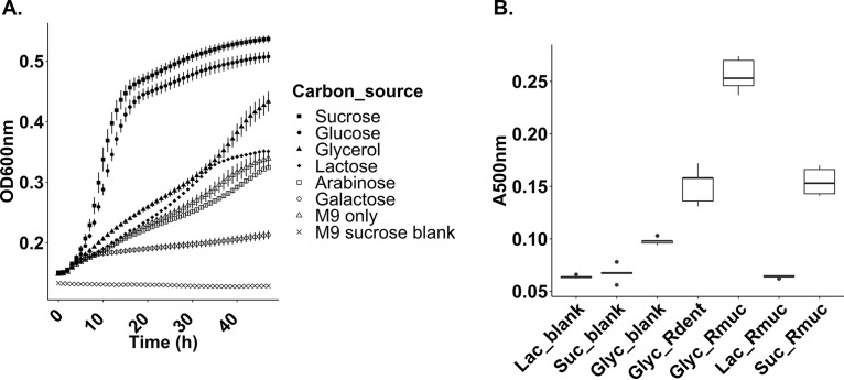
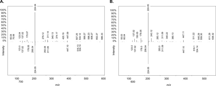
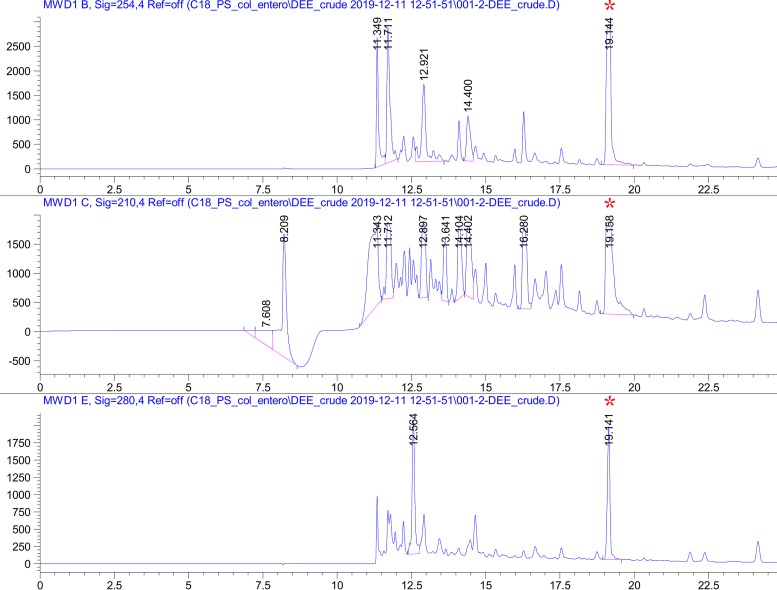
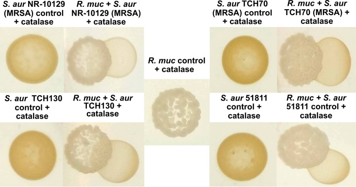
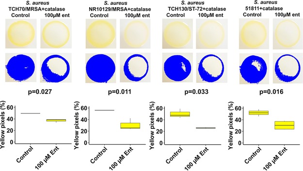
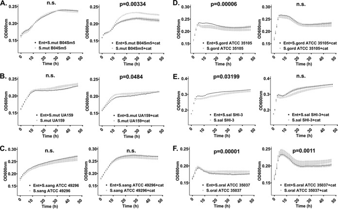
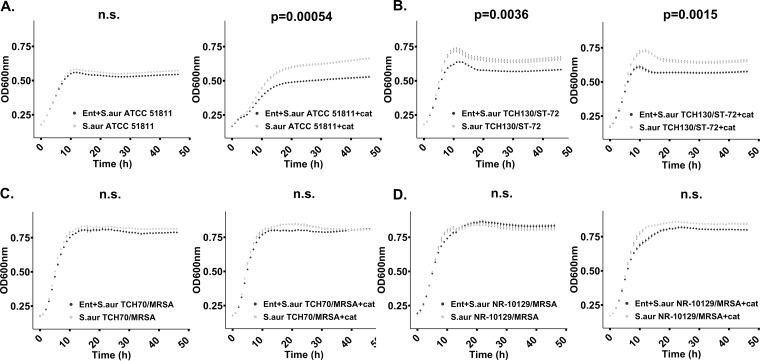
Next-generation sequencing studies of saliva and dental plaque from subjects in both healthy and diseased states have identified bacteria belonging to the Rothia genus as ubiquitous members of the oral microbiota. To gain a deeper understanding of molecular mechanisms underlying the chemical ecology of this unexplored group, we applied a genome mining approach that targets functionally important biosynthetic gene clusters (BGCs). All 45 genomes that were mined, representing Rothia mucilaginosa, Rothia dentocariosa, and Rothia aeria, harbored a catechol-siderophore-like BGC. To explore siderophore production further, we grew the previously characterized R. mucilaginosa ATCC 25296 in liquid cultures, amended with glycerol, which led to the identification of the archetype siderophore enterobactin by using tandem liquid chromatography-mass spectrometry (LC-MS/MS), high-performance liquid chromatography (HPLC), and nuclear magnetic resonance (NMR) spectroscopy. Normally attributed to pathogenic gut bacteria, R. mucilaginosa is the first commensal oral bacterium found to produce enterobactin. Cocultivation studies including R. mucilaginosa or purified enterobactin revealed that enterobactin reduced growth of certain strains of cariogenic Streptococcus mutans and pathogenic strains of Staphylococcus aureus Commensal oral bacteria were either unaffected, reduced in growth, or induced to grow adjacent to enterobactin-producing R. mucilaginosa or the pure compound. Taken together with Rothia's known capacity to ferment a variety of carbohydrates and amino acids, our findings of enterobactin production add an additional level of explanation to R. mucilaginosa's prevalence in the oral cavity. Enterobactin is the strongest Fe(III) binding siderophore known, and its role in oral health requires further investigation.IMPORTANCE The communication language of the human oral microbiota is vastly underexplored. However, a few studies have shown that specialized small molecules encoded by BGCs have critical roles such as in colonization resistance against pathogens and quorum sensing. Here, by using a genome mining approach in combination with compound screening of growth cultures, we identified that the commensal oral community member R. mucilaginosa harbors a catecholate-siderophore BGC, which is responsible for the biosynthesis of enterobactin. The iron-scavenging role of enterobactin is known to have positive effects on the host's iron pool and negative effects on host immune function; however, its role in oral health remains unexplored. R. mucilaginosa was previously identified as an abundant community member in cystic fibrosis, where bacterial iron cycling plays a major role in virulence development. With respect to iron's broad biological importance, iron-chelating enterobactin may explain R. mucilaginosa's colonization success in both health and disease.

Keywords: Actinomyces timonensis; Rothia mucilaginosa; Staphylococcus aureus; Streptococcus; Streptococcus spp.; enterobactin; oral microbiota.


Figures

FIG 1
FIG 1
(A) Growth curves for Rothia mucilaginosa ATCC 25296 incubated under aerobic conditions in liquid M9 medium, supplemented with different carbon sources (x axis, hours; y axis, optical density [OD600]). (B) Colorimetric absorbance at 500 nm (y axis, A500) capturing catecholate derivatives in liquid R. mucilaginosa (Rmuc) and R. dentocariosa M567 (Rdent) growth cultures (x axis) using Arnow’s assay. R. mucilaginosa was grown in M9 medium supplemented with 100 mM glycerol (Glyc_Rmuc), 100 mM lactate (Lac_Rmuc), and 100 mM sucrose (Suc_Rmuc). R. dentocariosa was grown in 100 mM glycerol to see if glycerol induced siderophore production as seen for R. mucilaginosa cultures.
FIG 2
FIG 2
Replicate mirror mass fragmentation patterns for enterobactin (m/z 670.152) produced by Rothia mucilaginosa ATCC 25296 (ion fragments pointing up) and enterobactin from the gold standard spectrum (ion fragments pointing down) in the Global Natural Products Social Molecular Networking (GNPS) library (36). Six major fragments (m/z 123.04, m/z 137.02, m/z 224.05, m/z 311.09 to 311.14, m/z 447.09 to 447.11, and m/z 534.14) of the query compound matched the gold standard in GNPS. (A) Purified extract derived from R. mucilaginosa ATCC 25296 growth medium (negative ionization mode). (B) Further enrichment of the siderophore using thin-layer chromatography (positive ionization mode). Both experiments confirmed the production of enterobactin.
FIG 3
FIG 3
High-performance liquid chromatography (HPLC) purification traces of enterobactin from Rothia mucilaginosa ATCC 25296 crude growth extract, measured at 210 nm, 254 nm, and 280 nm using a solvent gradient from 30 to 65% buffer B. The peak at 19.141 min (red asterisk) was eluted and further analyzed by NMR. x axis, minutes; y axis, peak absorbance intensity.
FIG 4
FIG 4
Rothia mucilaginosa ATCC 25296 (R. muc) inhibits pigment production in Staphylococcus aureus NR-10129 (MRSA), S. aureus TCH70 (MRSA), S. aureus TCH130, and enterotoxin H-producing S. aureus ATCC 51811, on M9 agar plates (100 mM glycerol) with 8 μg/ml catalase added. All S. aureus strains show yellow pigmentation when growing alone.
FIG 5
FIG 5
Yellow pigmentation of S. aureus strains exposed to 100 μM enterobactin and 8 μg/ml catalase on M9 agar plates supplemented with 100 μM glucose. Yellow pigmentation was measured with the R package “countcolors” (51). All strains presented statistically significant reductions in pigmentation in the presence of 100 μM enterobactin purified from R. mucilaginosa ATCC 25296 and catalase (P < 0.05, two-tailed t test). Box plots from yellow pixel measurements were generated with the R Studio program (49) and ggplot2 (50).
FIG 6
FIG 6
Growth curves of cariogenic and commensal Streptococcus species. Bacteria were grown aerobically at 37°C in liquid M9 medium supplemented with 100 mM glucose, 1 μM FeCl3, either with or without 100 μM enterobactin purified from R. mucilaginosa, and with or without 8 μg/ml catalase. Growth curves in black represent cultures amended with enterobactin. Statistically significant growth curves (P < 0.05) are shown with corresponding P values. Error bars reflect the standard error of the mean (calculated from triplicates). n.s., not significant. (A) Growth of cariogenic S. mutans strain B04Sm5 with enterobactin only (left panel) and with enterobactin and catalase (right panel). (B) Growth of cariogenic S. mutans strain UA159 with enterobactin only (left panel) and with enterobactin and catalase (right panel). (C) Growth of commensal S. sanguinis ATCC 49296 with enterobactin only (left panel) and with enterobactin and catalase (right panel). (D) Growth of S. gordonii ATCC 35101 with enterobactin only (left panel) and with enterobactin and catalase (right panel). (E) S. salivarius strain SHI-3 with enterobactin only (left panel) and with enterobactin and catalase (right panel). (F) Growth of S. oralis ATCC 35037 with enterobactin only (left panel) and with enterobactin and catalase (right panel). Graphs were generated and statistically validated using R Studio and the “statmod” and “ggplot2” packages (48–50).
FIG 7
FIG 7
Reduced growth of Staphylococcus aureus incubated in liquid M9 growth medium (100 μM enterobactin, 8 μg/ml catalase, 100 mM glucose) at 37°C for 24 h. No enterobactin was added to the control samples. Growth curves in black represent cultures amended with enterobactin. Statistically significant growth curves (P < 0.05) are shown with corresponding P values. Error bars reflect the standard error of the mean (calculated from triplicates). (A) Growth of S. aureus strain 51811 with enterobactin only (left panel) and with enterobactin and catalase (right panel). (B) Growth of S. aureus strain TCH130/ST-72 with enterobactin only (left panel) and with enterobactin and catalase (right panel). (C) Growth of S. aureus TCH70/MRSA with enterobactin only (left panel) and with enterobactin and catalase (right panel). (D) Growth of S. aureus NR-10129/MRSA with enterobactin only (left panel) and with enterobactin and catalase (right panel). Graphs were generated and statistically validated using R Studio and the “statmod” and “ggplot2” packages (48–50).

References

    1. Lux T, Nuhn M, Hakenbeck R, Reichmann P. 2007. Diversity of bacteriocins and activity spectrum in Streptococcus pneumoniae. J Bacteriol 189:7741–7751. doi:10.1128/JB.00474-07. - DOI - PMC - PubMed
    1. Nes IF, Diep DB, Holo H. 2007. Bacteriocin diversity in Streptococcus and Enterococcus. J Bacteriol 189:1189–1198. doi:10.1128/JB.01254-06. - DOI - PMC - PubMed
    1. Tang X, Kudo Y, Baker JL, LaBonte S, Jordan PA, McKinnie SMK, Guo J, Huan T, Moore BS, Edlund A. 2020. Cariogenic Streptococcus mutans produces tetramic acid strain-specific antibiotics that impair commensal colonization. ACS Infect Dis 6:563–571. doi:10.1021/acsinfecdis.9b00365. - DOI - PMC - PubMed
    1. Aleti G, Baker JL, Tang X, Alvarez R, Dinis M, Tran NC, Melnik AV, Zhong C, Ernst M, Dorrestein P, Edlund A. 2019. Identification of the bacterial biosynthetic gene clusters of the oral microbiome illuminates the unexplored social language of bacteria during health and disease. mBio 10:e00321-19. doi:10.1128/mBio.00321-19. - DOI - PMC - PubMed
    1. Donia MS, Cimermancic P, Schulze CJ, Wieland Brown LC, Martin J, Mitreva M, Clardy J, Linington RG, Fischbac MA. 2014. A systematic analysis of biosynthetic gene clusters in the human microbiome reveals a common family of antibiotics. Cell 158:1402–1414. doi:10.1016/j.cell.2014.08.032. - DOI - PMC - PubMed