. 2020 Apr 28;10(1):7103.
doi: 10.1038/s41598-020-63183-5.

Investigating APOE, APP-Aβ metabolism genes and Alzheimer's disease GWAS hits in brain small vessel ischemic disease

Affiliations

Investigating APOE, APP-Aβ metabolism genes and Alzheimer's disease GWAS hits in brain small vessel ischemic disease

Sonja Blumenau et al. Sci Rep. .

Abstract

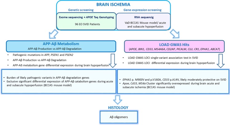
Alzheimer's disease and small vessel ischemic disease frequently co-exist in the aging brain. However, pathogenic links between these 2 disorders are yet to be identified. Therefore we used Taqman genotyping, exome and RNA sequencing to investigate Alzheimer's disease known pathogenic variants and pathways: APOE ε4 allele, APP-Aβ metabolism and late-onset Alzheimer's disease main genome-wide association loci (APOE, BIN1, CD33, MS4A6A, CD2AP, PICALM, CLU, CR1, EPHA1, ABCA7) in 96 early-onset small vessel ischemic disease Caucasian patients and 368 elderly neuropathologically proven controls (HEX database) and in a mouse model of cerebral hypoperfusion. Only a minority of patients (29%) carried APOE ε4 allele. We did not detect any pathogenic mutation in APP, PSEN1 and PSEN2 and report a burden of truncating mutations in APP-Aß degradation genes. The single-variant association test identified 3 common variants with a likely protective effect on small vessel ischemic disease (0.54>OR > 0.32, adj. p-value <0.05) (EPHA1 p.M900V and p.V160A and CD33 p.A14V). Moreover, 5/17 APP-Aß catabolism genes were significantly upregulated (LogFC > 1, adj. p-val<0.05) together with Apoe, Ms4a cluster and Cd33 during brain hypoperfusion and their overexpression correlated with the ischemic lesion size. Finally, the detection of Aβ oligomers in the hypoperfused hippocampus supported the link between brain ischemia and Alzheimer's disease pathology.


Conflict of interest statement

The authors declare no competing interests.

Figures

Figure 1
Figure 1
Pipeline followed in the study. SVID, small vessel ischemic disease; VaD, vascular dementia; BCCAS, bilateral common carotid artery stenosis; LOAD, late-onset Alzheimer’s disease; GWAS, genome-wide association study.
Figure 2
Figure 2
Number of loss of function (LoF) mutations in APP-Aβ degradation and production genes detected in the HEX and SVID cohorts and number of individuals per each cohort. The SVID cohort presents a burden of truncating mutations, compared to the HEX cohort. SVID, small vessel ischemic disease.
Figure 3
Figure 3
A-G.Bilateral common carotid artery stenosis (BCCAS) mouse model, displaying monolateral right small subcortical lesions, mainly affecting striatum (A) and hippocampus (B) (blue arrows), during acute (2d) and subacute (7d) hypoperfusion. The most severly hypoperfused hemisphere, with ischemic lesions detected on T2 weighted MRI (right hemisphere) was used for RNA sequencing. The contralateral hypoperfused hemisphere, with no ischemic lesions detectable on T2-MRI (left hemisphere) was used for immunohistochemistry. C, hypoperfused brain stained with GFAP, presenting peri-infarct astrocytosis in hippocampus and striatum during subcute hypoperfusion (7d) (white arrows). The infarct area is delimited by white dashed lines. The left hemisphere is hypoperfused and although ischemic lesions in the left side are not detectable on T2-MRI, we report a significant gliosis (yellow arrow). D, hypoperfused brain stained with IBA1, presenting microglia infiltration of the infarct areas and to a lesser extent peri-infarct areas and in the most severe hypoperfused areas during subcute hypoperfusion (7d) (pink arrows). E. Hypoperfused hippocampus stained with GFAP, displaying astrocytosis. F. Hypoperfused hippocampus stained with IBA1, displaying gliosis. G. T2-MRI slides showing the extension of the ischemic lesion (red).
Figure 4
Figure 4
(A) Cerebral blood flow (CBF) reduction detected on MRI in hippocampus during acute (2d) and subacute (7d) hypoperfusion in BCCAS mice. The CBF drop at day 1 reaches 60–70% of the CBF values detected before the surgery (d0) or in naive mice and progressively recovers.CBF, cerebral blood flow; d, day; BCCAS, bilateral common carotid artery stenosis. (B) Differential gene expression, expressed in transcript per million (TPM) in APP-Aß metabolism genes and LOAD GWAS loci detected in BCCAS mice in hippocampus during subacute hypoperfusion (7d). C Percentage of ischemic lesion volume detected on T2-MRI in hippocampus of BCCAS mice during subacute hypoperfusion. H,hippocampus; L, left; R, right; d, day.
Figure 5
Figure 5
Toxic Aβ oligomers are detected in hypoperfused hippocampus mostly in the CA1 region during subacute hypoperfusion (7d) (B,B’) and absent in naive hippocampus (A,A’). The Aβ oligomers co-localize with reactive astrocytes (C-D). The number of glial and neuronal cells positive for Aβ oligomers are significantly higher in hippocampus during subacute hypoperfusion (7d) compared to acute hypoperfusion (2d) and naive mice (E). BCCAS, bilateral common carotid artery stenosis; d, day; +, positive.

References

    1. Lim A, et al. Clinico-neuropathological correlation of Alzheimer’s disease in a community-based case series. J Am Geriatr Soc. 1999;47:564–569. doi: 10.1111/j.1532-5415.1999.tb02571.x. - DOI - PubMed
    1. Langa KM, Foster NL, Larson EB. Mixed dementia: emerging concepts and therapeutic implications. JAMA. 2004;292:2901–2908. doi: 10.1001/jama.292.23.2901. - DOI - PubMed
    1. Corder EH, et al. Gene dose of apolipoprotein E type 4 allele and the risk of Alzheimer’s disease in late onset families. Science. 1993;261:921–923. doi: 10.1126/science.8346443. - DOI - PubMed
    1. Bu G. Apolipoprotein E and its receptors in Alzheimer’s disease: pathways, pathogenesis and therapy. Nat. Rev. Neurosci. 2009;10:333–344. doi: 10.1038/nrn2620. - DOI - PMC - PubMed
    1. Huang Y, Mucke L. Alzheimer mechanisms and therapeutic strategies. Cell. 2012;148:1204–1222. doi: 10.1016/j.cell.2012.02.040. - DOI - PMC - PubMed

Publication types

MeSH terms