. 2020 Apr 28;10(1):7122.
doi: 10.1038/s41598-020-63956-y.

The internal cranial anatomy of Champsosaurus (Choristodera: Champsosauridae): Implications for neurosensory function

Affiliations

The internal cranial anatomy of Champsosaurus (Choristodera: Champsosauridae): Implications for neurosensory function

Thomas W Dudgeon et al. Sci Rep. .

Abstract

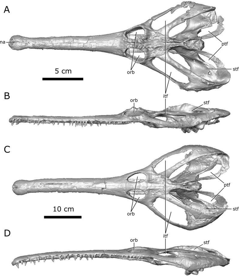
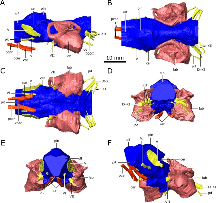
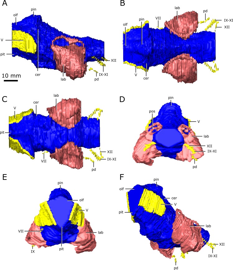
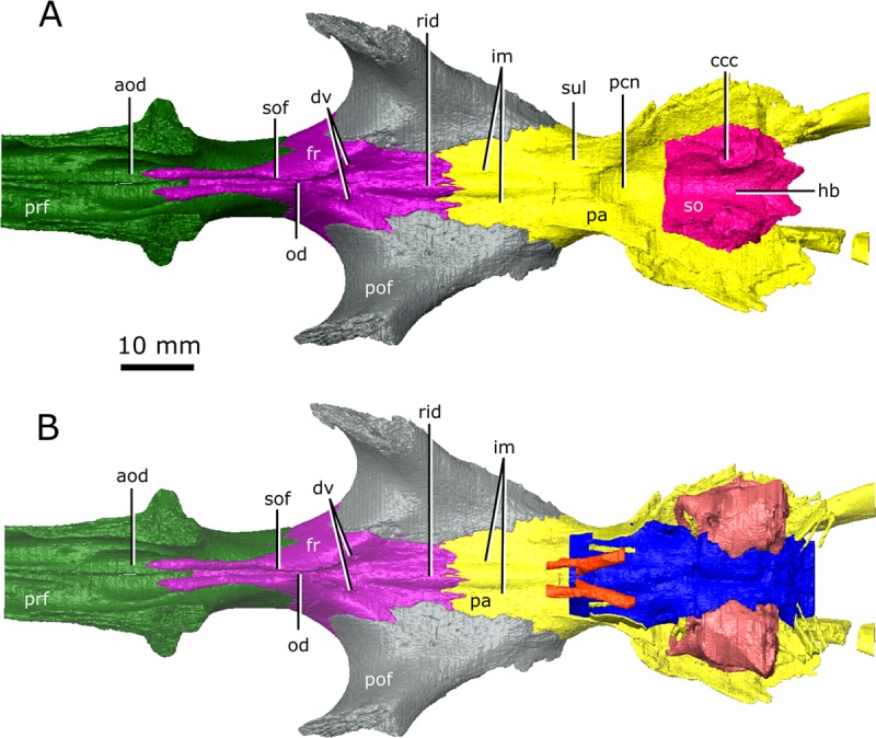
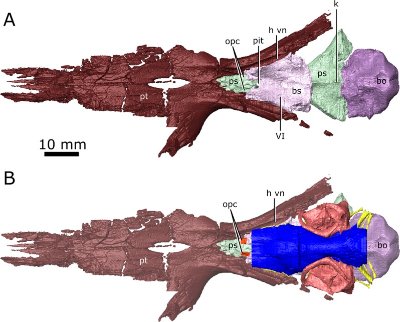
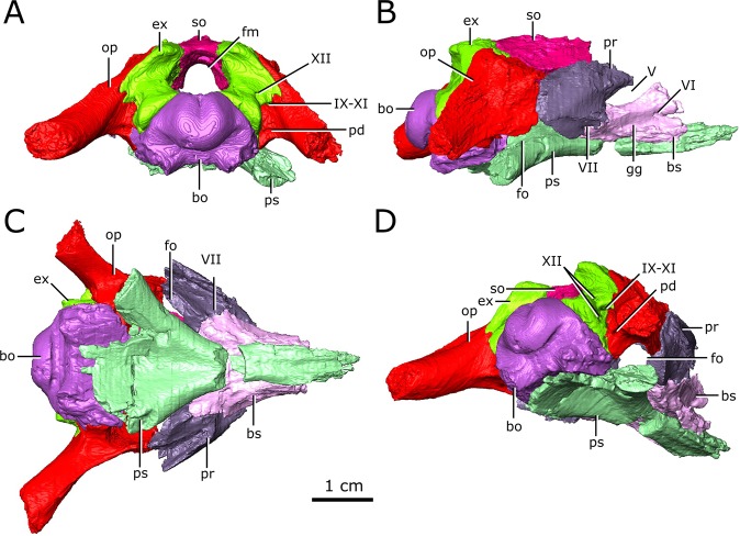
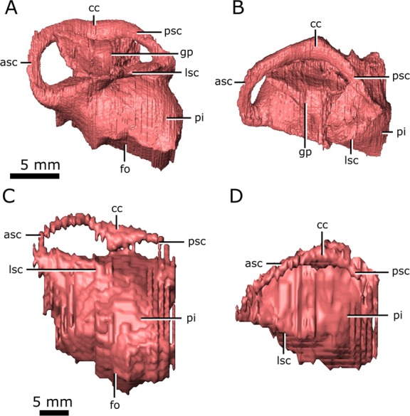
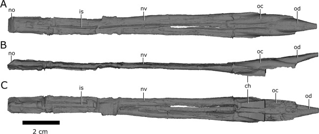
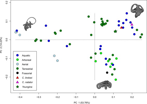
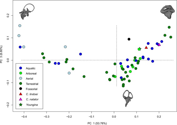
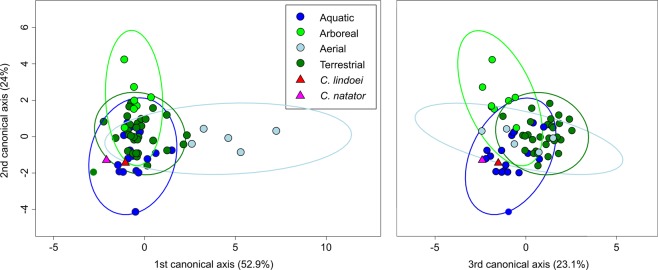
Although isolated Champsosaurus remains are common in Upper Cretaceous sediments of North America, the braincase of these animals is enigmatic due to the fragility of their skulls. Here, two well-preserved specimens of Champsosaurus (CMN 8920 and CMN 8919) are CT scanned to describe their neurosensory structures and infer sensory capability. The anterior portion of the braincase was poorly ossified and thus does not permit visualization of a complete endocast; however, impressions of the olfactory stalks indicate that they were elongate and likely facilitated good olfaction. The posterior portion of the braincase is ossified and morphologically similar to that of other extinct diapsids. The absence of an otic notch and an expansion of the pars inferior of the inner ear suggests Champsosaurus was limited to detecting low frequency sounds. Comparison of the shapes of semicircular canals with lepidosaurs and archosauromorphs demonstrates that the semicircular canals of Champsosaurus are most similar to those of aquatic reptiles, suggesting that Champsosaurus was well adapted for sensing movement in an aquatic environment. This analysis also demonstrates that birds, non-avian archosauromorphs, and lepidosaurs possess significantly different canal morphologies, and represents the first morphometric analysis of semicircular canals across Diapsida.


Conflict of interest statement

The authors declare no competing interests.

Figures

Figure 1
Figure 1
Digitized models of the skulls of Champsosaurus lindoei (CMN 8920) and Champsosaurus natator (CMN 8919) based on micro-computed tomography scanning. (A) CMN 8920 in dorsal view; (B) CMN 8920 in left lateral view; (C) CMN 8919 in dorsal view; (D) CMN 8919 in left lateral view. Abbreviations: itf, infratemporal fenestra; na, narial opening; orb, orbit; ptf, post-temporal fenestra; stf, supratemporal fenestra. Images generated in Amira 5.4.3 (https://www.fei.com/software/amira/) and processed in Inkscape 0.92 (https://inkscape.org/).
Figure 2
Figure 2
Reconstruction of the endocranial anatomy of Champsosaurus lindoei (CMN 8920). (A) left lateral view; (B) dorsal view; (C) ventral view; (D) posterior view; (E) anterior view; (F) left anterolateroventral view. The brain endocast is illustrated in blue, endosseous labyrinth in pink, cranial nerves in yellow, and carotid artery in red. Abbreviations: car, carotid arteries; ccar, cerebral branch of the carotid arteries; cer, cerebrum; lab, endosseous labyrinth; olf, base of the olfactory lobes; pcar, palatine branch of the carotid arteries; pd, canal for the parilymphatic duct; pin, pineal body; pit, pituitary fossa; IX-XI, canal for cranial nerves IX, X, and XI; XII, canal for cranial nerve XII; V, opening for cranial nerve V; VI, canal for cranial nerve VI; VII, canal for cranial nerve VII. Images generated in Amira 5.4.3 (https://www.fei.com/software/amira/) and processed in Inkscape 0.92 (https://inkscape.org/).
Figure 3
Figure 3
Reconstruction of the endocranial anatomy of Champsosaurus natator (CMN 8919). (A) left lateral view; (B) dorsal view; (C) ventral view; (D) posterior view; (E) anterior view; (F) left anterolateroventral view. The brain endocast is illustrated in blue, endosseous labyrinth in pink, cranial nerves in yellow, and carotid artery in red. Abbreviations: cer, cerebrum; lab, endosseous labyrinth; olf, base of the olfactory lobes; pd, canal for the parilymphatic duct; pin, pineal body; pit, pituitary fossa; IX-XI, canal for cranial nerves IX, X, and XI; XII, canal for cranial nerve XII; V, opening for cranial nerve V; VII, canal for cranial nerve VII. Images generated in Amira 5.4.3 (https://www.fei.com/software/amira/) and processed in Inkscape 0.92 (https://inkscape.org/).
Figure 4
Figure 4
The isolated braincase roof (prefrontals, frontals, postfrontals, parietals, supraoccipital) of CMN 8920. (A) Ventral view of the isolated braincase roof; (B) Ventral view of the braincase roof with the segmented brain endocast (blue), endosseous labyrinth (pink), cranial nerves (yellow), and carotid arteries (red) in position. The braincase roof is slightly faded. Abbreviations: aod, anterior of olfactory duct; ccc, canal for the crus communis; dv, diploic vein foramen; fr, frontal; hb, roof of the hindbrain; im, impressions of the olfactory tracks; od, olfactory duct; pcn, parietal concavity for the pineal body; pa, parietal; pof, postfrontal; prf, prefrontal; rid, ridge seperating the paired olfactory tracts; so, supraoccipital; sof, subolfactory flange; sul, area inundated with sulci. Images generated in Amira 5.4.3 (https://www.fei.com/software/amira/) and processed in Inkscape 0.92 (https://inkscape.org/).
Figure 5
Figure 5
The isolated braincase floor (pterygoids, basisphenoid, parasphenoid, and basioccipital) of CMN 8920. (A) Dorsal view of the isolated braincase floor; (B) Dorsal view of the braincase floor with the segmented brain endocast (blue), endosseous labyrinth (pink), cranial nerves (yellow), and carotid arteries (red) in position. The braincase floor is slightly faded. Abbreviations: bo, basioccipital; bs, basisphenoid; hvn, trough for the lateral head vein; k, parasphenoid keel; opc, opening for the palatine branch of the carotid artieries; pit, pituitary fossa; ps, parasphenoid; pt, pterygoid; VI, exit for cranial nerve VI. Images generated in Amira 5.4.3 (https://www.fei.com/software/amira/) and processed in Inkscape 0.92 (https://inkscape.org/).
Figure 6
Figure 6
Isolated chondrocranial elements and parasphenoid of CMN 8920. (A) posterior view; (B) right lateral view; (C) ventral view; (D) posterolateroventral view. Abbreviations: bo, basioccipital; bs basisphenoid; ex, exoccipital; fm, foramen magnum; fo, fenestra ovalis; gg, depression for the gasserian ganglion; op, opisthotic; pd, foramen for the perilymphatic duct; pr, prootic; ps, parasphenoid; so, supraoccipital; Roman numerals indicate the foramina of the corresponding cranial nerves. Images generated in Amira 5.4.3 (https://www.fei.com/software/amira/) and processed in Inkscape 0.92 (https://inkscape.org/).
Figure 7
Figure 7
Left endosseous labyrinth of Champsosaurus lindoei (CMN 8920) in: (A) lateral view; and (B) dorsal view, and Champsosaurus natator (CMN 8919) in: (C) lateral view; and (D) dorsal view. Abbreviations: asc, anterior semicircular canal; cc, crus communis, fo, fenestra ovalis; gp, unossified gap between the prootic, opisthotic, and supraoccipital; lsc, lateral semicircular canal; pi, pars inferior; psc, posterior semicircular canal. Images generated in Amira 5.4.3 (https://www.fei.com/software/amira/) and processed in Inkscape 0.92 (https://inkscape.org/).
Figure 8
Figure 8
Left and right nasal passages of Champsosaurus lindoei (CMN 8920), in (A) dorsal view; (B) left lateral view; and (C) ventral view. Abbreviations: ch, choana; is, ridge indicating the internarial septum; no, narial opening; nv, nasal vestibule; oc, olfactory chamber; od, olfactory duct. Images generated in Amira 5.4.3 (https://www.fei.com/software/amira/) and processed in Inkscape 0.92 (https://inkscape.org/).
Figure 9
Figure 9
Correlation between scaled and transformed endocochlear duct length (ECD) and (A) mean best hearing frequency (y = 3391.3x + 4026.8; r2 = 0.5825; p = 2.28e-05); (B) best hearing range (y = 6190x + 7003.193; r2 = 0.5521; p = 4.875e-05) with ecologies coloured separately. Grey area indicates the 95% confidence interval of the regression line. Extant data from Walsh et al. (2009). Red triangle indicates the predicted value for Champsosaurus lindoei (CMN 8920).
Figure 10
Figure 10
PC 1 vs PC 2, representing 67.98% of the total variation. Taxa are colour coded based on ecology. End-point morphologies: top right, Tomistoma schlegelii; bottom, Aheatulla nasuta; top left, Passer domesticus.
Figure 11
Figure 11
PC 1 vs PC 3 representing 60.56% of the total variation. Taxa are colour coded based on ecology. End-point morphologies: top left, Passer domesticus; bottom, Erlikosaurus andrewsi; top right, Manouria emys.
Figure 12
Figure 12
CV1 vs CV2 (left) representing 76.9% of the total between-group variation, and CV3 vs CV2 (right) representing 47.1% of the total between-group cariation. 95% confidence ellipses of each ecological group are plotted.

References

    1. Witmer, L. M., Ridgely, R. C., Dufeau, D. L., & Semones, M. C. Using CT to peer into the past: 3D visualization of the brain and ear regions of birds, crocodiles, and nonavian dinosaurs. In Anatomical imaging: Towards a new morphology (eds. Endo, H., & Frey, R.) 65-87 (Springer, 2008).
    1. Jerison, H. J. Evolution of the brain and intelligence. (Academic Press, 1973).
    1. Alonso PD, Milner AC, Ketcham RA, Cookson MJ, Rowe TB. The avian nature of the brain and inner ear of Archaeopteryx. Nature. 2004;430:666–669. doi: 10.1038/nature02706. - DOI - PubMed
    1. Witmer LM, Ridgely RC. New insights into the brain, braincase, and ear region of tyrannosaurs (Dinosauria, Theropoda), with implications for sensory organization and behavior. Anat. Rec. 2009;292:1266–1296. doi: 10.1002/ar.20983. - DOI - PubMed
    1. Evans DC. New evidence on brain−endocranial cavity relationships in ornithischian dinosaurs. Acta Palaeontol. Pol. 2005;50:617–622.

Publication types