. 2020 Apr 15:16:977-984.
doi: 10.2147/NDT.S236939. eCollection 2020.

Clinical, Electrophysiological and Radiological Features of Nitrous Oxide-Induced Neurological Disorders

Affiliations

Clinical, Electrophysiological and Radiological Features of Nitrous Oxide-Induced Neurological Disorders

Lei Bao et al. Neuropsychiatr Dis Treat. .

Abstract

Purpose: We summarized the clinical manifestations, laboratory and electrodiagnostic characteristics and magnetic resonance imaging (MRI) findings of nitrous oxide (N2O) abuse-induced neurological disorders.

Patients and methods: We retrospectively reviewed 33 patients with N2O abuse-induced neurological disorders and reported their demographic data, clinical manifestations, laboratory examinations, nerve conduction studies, together with spinal and brain MRI.

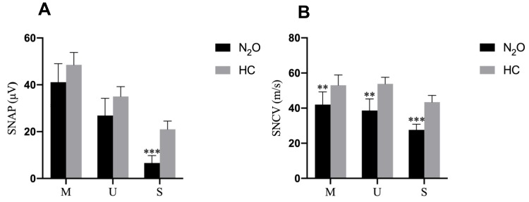
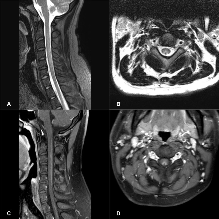
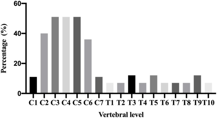
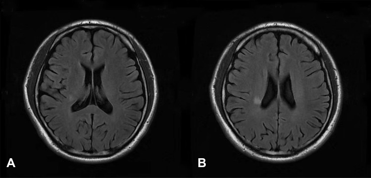
Results: The most frequent clinical manifestations included numbness and weakness in the extremities and unspecified gait disturbance. Low serum vitamin B12 levels were found in 9 patients, and high homocysteine levels were noted in 27 patients. Nerve conduction studies showed a sensory-motor neuropathy. Sixteen patients showed bilateral high-intensity T2 signal within the posterior column on spinal MRI, and four patients showed cerebral white matter lesions on brain MRI.

Conclusion: N2O abuse has become a significant public health problem because of the severe neurological disorders related to chronic abuse. Clinical physicians should be aware of the toxic effects of N2O.

Keywords: neurological disorders; neuropathy; nitrous oxide; subacute combined degeneration; vitamin B12.


Conflict of interest statement

The authors declare no competing financial interests.

Figures

Figure 1
Figure 1
Motor nerve conduction study results in the health control (HC) and N2O abuse groups. (A) The compound muscle action potential amplitude, (B) motor conduction velocity, (C) distal latency, and (D) F-wave latency of each motor nerve for the HC (grey bar) and N2O-abuse (black bar) groups. Notes: Data are presented as mean ± standard error of the mean. Significant difference is indicated by **p < 0.01; ***p < 0.001. Abbreviations: M, median nerve; U, ulnar nerve; P, peroneal nerve; T, tibial nerve.
Figure 2
Figure 2
Sensory nerve conduction study results in the health control (HC) and N2O-abuse groups. (A) The sensory nerve action potential amplitude and (B) sensory conduction velocity of each sensory nerve for the HC (gray bar) and N2O-abuse (black bar) groups. Notes: Data are presented as mean ± standard error of the mean. Significant differences are indicated by **p < 0.01; ***p < 0.001. Abbreviations: M, median nerve; U, ulnar nerve; S, sural nerve.
Figure 3
Figure 3
Spinal cord MRI of a N2O-abuse patient who complained of unsteadiness while walking and limb numbness for 2 weeks. (A) Sagittal T2 MRI of the cervical spine demonstrating a high-signal lesion representing demyelination C2-C6. (B) Axial T2 MRI of the cervical spine shows hyperintense, symmetric and inverted V-shaped signals involving the dorsal columns of C2-C6. (C) Gadolinium-enhanced T1 MRI reveals the enhancement of the dorsal columns. (D) Gadolinium-enhanced axial T1 MRI shows a V-shaped enhancement of the dorsal columns.
Figure 4
Figure 4
The distribution of lesions detected in the spinal cord of N2O-abuse patients. The cervical spinal cord was more frequently impaired than other areas of the spinal cord. C3, C4 and C5 were the most common affected vertebral level.
Figure 5
Figure 5
Axial fluid-attenuated inversion recovery (FLAIR) scan of a N2O-abused patient who complained of memory decline for 1 month. (A and B) showed mild white matter lesions in periventricular regions.

References

    1. Nagele P, Duma A, Kopec M, et al. Nitrous oxide for treatment-resistant major depression: a proof-of-concept trIal. Biol Psychiatry. 2015;78(1):10–18. doi:10.1016/j.biopsych.2014.11.016 - DOI - PubMed
    1. Sanders RD, Weimann J, Maze M. Biologic effects of nitrous oxide: a mechanistic and toxicologic review. Anesthesiology. 2008;109(4):707–722. doi:10.1097/ALN.0b013e3181870a17 - DOI - PubMed
    1. van Amsterdam J, Nabben T, van den Brink W. Recreational nitrous oxide use: prevalence and risks. Regul Toxicol Pharmacol. 2015;73(3):790–796. doi:10.1016/j.yrtph.2015.10.017 - DOI - PubMed
    1. Lan SY, Kuo CY, Chou CC, et al. Recreational nitrous oxide abuse related subacute combined degeneration of the spinal cord in adolescents - A case series and literature review. Brain Dev. 2019;41(5):428–435. doi:10.1016/j.braindev.2018.12.003 - DOI - PubMed
    1. Oussalah A, Julien M, Levy J, et al. Global burden related to nitrous oxide exposure in medical and recreational settings: a systematic review and individual patient data meta-analysis. J Clin Med. 2019;8:4. doi:10.3390/jcm8040551 - DOI - PMC - PubMed