. 2020 Aug;10(4):320-328.
doi: 10.1016/j.jpha.2020.04.008. Epub 2020 Apr 28.

Structural elucidation of SARS-CoV-2 vital proteins: Computational methods reveal potential drug candidates against main protease, Nsp12 polymerase and Nsp13 helicase

Affiliations

Structural elucidation of SARS-CoV-2 vital proteins: Computational methods reveal potential drug candidates against main protease, Nsp12 polymerase and Nsp13 helicase

Muhammad Usman Mirza et al. J Pharm Anal. 2020 Aug.

Abstract

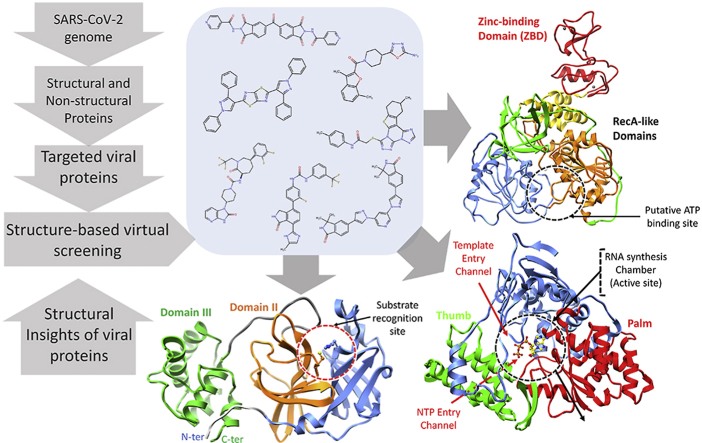
Recently emerged SARS-CoV-2 caused a major outbreak of coronavirus disease 2019 (COVID-19) and instigated a widespread fear, threatening global health safety. To date, no licensed antiviral drugs or vaccines are available against COVID-19 although several clinical trials are under way to test possible therapies. During this urgent situation, computational drug discovery methods provide an alternative to tiresome high-throughput screening, particularly in the hit-to-lead-optimization stage. Identification of small molecules that specifically target viral replication apparatus has indicated the highest potential towards antiviral drug discovery. In this work, we present potential compounds that specifically target SARS-CoV-2 vital proteins, including the main protease, Nsp12 RNA polymerase and Nsp13 helicase. An integrative virtual screening and molecular dynamics simulations approach has facilitated the identification of potential binding modes and favourable molecular interaction profile of corresponding compounds. Moreover, the identification of structurally important binding site residues in conserved motifs located inside the active site highlights relative importance of ligand binding based on residual energy decomposition analysis. Although the current study lacks experimental validation, the structural information obtained from this computational study has paved way for the design of targeted inhibitors to combat COVID-19 outbreak.

Keywords: COVID-19 outbreak; CoV-Mpro; CoV-Nsp12 polymerase; CoV-Nsp13 helicase; SARS-CoV-2.


Conflict of interest statement

The authors declare that there are no conflicts of interest.

Figures

Image 1
Graphical abstract
Fig. 1
Fig. 1
Structural representation of SARS-CoV-2 proteins. (A) SARS-CoV-2 main protease monomer (ribbon representation) composed of: (i) N-terminal domain I (cornflower blue), (ii) domain II (orange), and (iii) C-terminal domain III (green). Substrate recognition site in circle (red) and catalytic dyad residues, His41 and Cys145 are highlighted and labelled. (B) Linear schematic description of domain architecture of SARS-CoV-2 Nsp12 polymerase followed by its structure composed of thumb (green), palm (red), and finger (cornflower blue) subdomains. The active site of Nsp12 polymerase is highlighted and arrangement of structurally conserved RdRp motifs in Nsp12 polymerase model coloured green (A), yellow (B), pink (C), orange (D), brown (E), cornflower blue (F) and magenta (G) for motifs A-G respectively is displayed in bottom right. Superposition of the polio virus elongation complex structure (PDB: 3OL8) CTP (orange) inside the predicted binding site also displayed. (C) Overall structure of SARS-CoV-2 Nsp13 helicase composed of ZBD (red), stalk (golden), 1B (green), 1A (orange) and 2A (cornflower blue) domains. Three zinc atoms in ZBD are shown as dark grey spheres. The binding pocket residues are zoomed in and labelled. Linear schematic diagram of the domain organization of SARS-CoV-2 Nsp13 helicase is displayed at the bottom.
Fig. 2
Fig. 2
Post-molecular dynamics (MD) analysis of SARS-CoV-2 Mpro hits. (A) Molecular surface representation of Mpro with MD simulated representative conformation of cmp3 (yellow), cmp12 (green), cmp14 (magenta), cmp17 (cyan) and cmp18 (dark blue) inside the substrate binding site zoomed with subsites S1 (orange), S2 (pink) and S4 (cyan) and residues are labelled accordingly. Molecular interactions representations of each complex with interacting residues are highlighted in blue sticks and catalytic dyad in green sticks. (B) Per-residue energy decomposition analysis of potential substrate-binding site residues. Terminal moieties of cmp12 and cmp14 revealed similar binding modes while cmp3, cmp17 and cmp18 showing different binding mode are displayed in (C) and (D) followed by chemical structure representations of these compounds in connection with interactions of structural moieties with binding pocket subsites.
Fig. 3
Fig. 3
Post-molecular dynamics (MD) analysis of SARS-CoV-2 Nsp12 RdRp complexes. (A) MD simulated conformations of cmp2 (green) (B), cmp17a (orange) (C), cmp21 (magenta) (D) inside the predicted binding pocket of Nsp12 polymerase. The arrangement of motifs and colors are the same as in Fig. 1B. Molecular interactions of individual compounds are displayed in B and C and D, and residues are labelled accordingly. (E) Representations of chemical structures of compounds, and (F) Per-residue energy decomposition analysis of potential binding pocket residues.
Fig. 4
Fig. 4
Post-molecular dynamics (MD) analysis of SARS-CoV-2 Nsp13 helicase complexes. (A) Yeast Upf1-ADP complex (transparent white) superimposed on Nsp13 helicase to locate the ATP putative binding pocket and 2D-interaction plot is displayed. MD simulated cmp1 (brown), cmp3a (magenta), cmp11 (green) and cmp15 (sandy brown) are displayed inside the pocket. The arrangement of domains is coloured the same as in Fig. 1C. (B) MD simulated conformations of cmp11 (green) inside the predicted binding pocket of Nsp13 helicase interacted with key residues of domain 1A, 2A and 1B. (C) Per-residue energy decomposition analysis of potential binding pocket residues, and representations of chemical structures of compounds are displayed in (D).

References

    1. Benvenuto D., Giovanetti M., Ciccozzi A. bioRxiv; 2020. The 2019-new coronavirus epidemic: Evidence for virus evolution. - PMC - PubMed
    1. Zhu R.-f., Gao R.-l., Robert S.-H. medRxiv; 2020. Systematic review of the registered clinical trials of coronavirus diseases 2019 (COVID-19) - PMC - PubMed
    1. Yamamoto N., Matsuyama S., Hoshino T. bioRxiv; 2020. Nelfinavir inhibits replication of severe acute respiratory syndrome coronavirus 2 in vitro.
    1. Wang M., Cao R., Zhang L. Remdesivir and chloroquine effectively inhibit the recently emerged novel coronavirus (2019-nCoV) in vitro. Cell Res. 2020;30:269–271. - PMC - PubMed
    1. Holshue M.L., DeBolt C., Lindquist S. First case of 2019 novel coronavirus in the United States. N. Engl. J. Med. 2020;382:929–936. - PMC - PubMed