

Coronavirus disease 2019 (COVID-19) imaging reporting and data system (COVID-RADS) and common lexicon: a proposal based on the imaging data of 37 studies
- PMID: 32346790
- PMCID: PMC7186323
- DOI: 10.1007/s00330-020-06863-0
Coronavirus disease 2019 (COVID-19) imaging reporting and data system (COVID-RADS) and common lexicon: a proposal based on the imaging data of 37 studies
Abstract
Background: In the vast majority of the laboratory-confirmed coronavirus disease 2019 (COVID-19) patients, computed tomography (CT) examinations yield a typical pattern and the sensitivity of this modality has been reported to be 97% in a large-scale study. Structured reporting systems simplify the interpretation and reporting of imaging examinations, serve as a framework for consistent generation of recommendations, and improve the quality of patient care.
Purpose: To compose a comprehensive lexicon for description of the imaging findings and propose a grading system and structured reporting format for CT findings in COVID-19.
Material and methods: We updated our published systematic review on imaging findings in COVID-19 to include 37 published studies pertaining to diagnostic features of COVID-19 in chest CT. Using the reported imaging findings of 3647 patients, we summarized the typical chest CT findings, atypical features, and temporal changes of COVID-19 in chest CT. Subsequently, we extracted a list of descriptive terms and mapped it to the terminology that is commonly used in imaging literature.
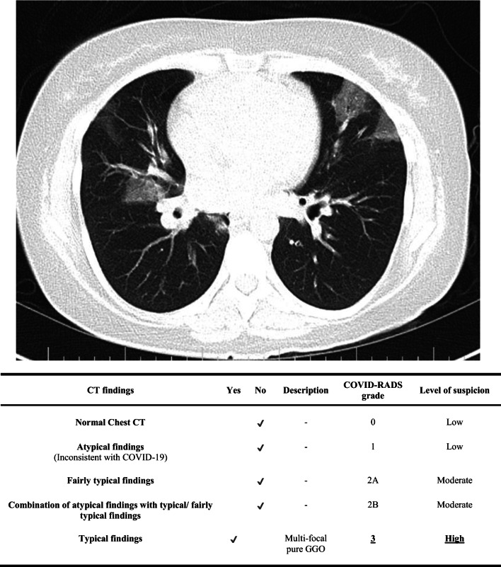
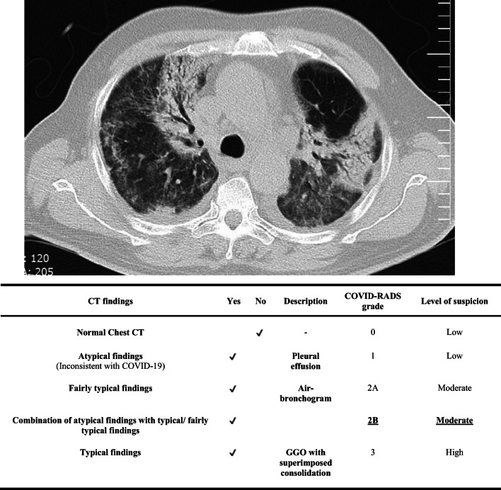
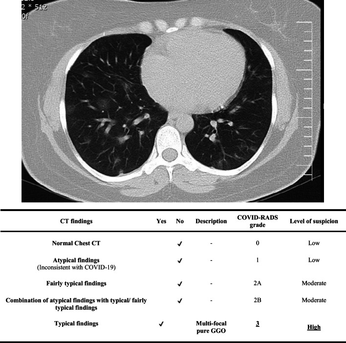
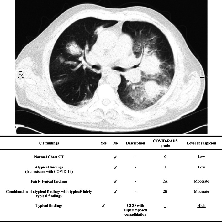
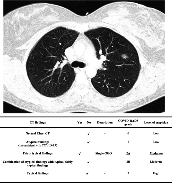
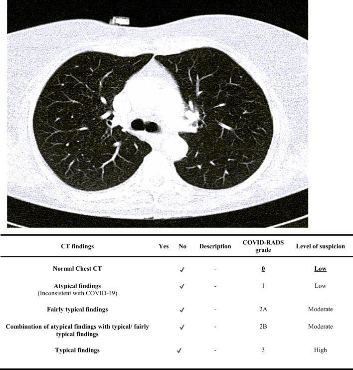
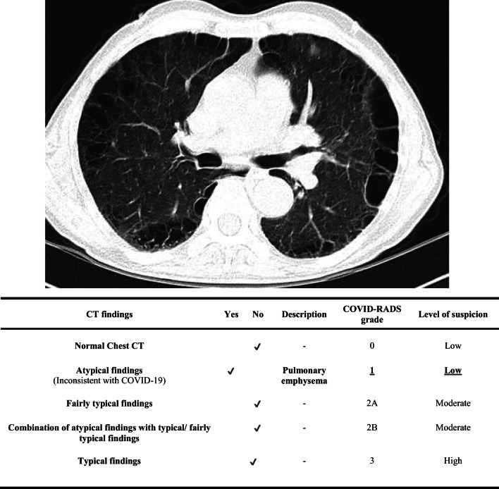
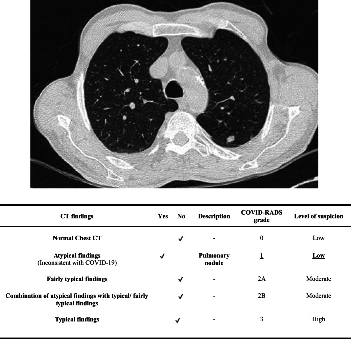
Results: We composed a comprehensive lexicon that can be used for documentation and reporting of typical and atypical CT imaging findings in COVID-19 patients. Using the same data, we propose a grading system with five COVID-RADS categories. Each COVID-RADS grade corresponds to a low, moderate, or high level of suspicion for pulmonary involvement of COVID-19.
Conclusion: The proposed COVID-RADS and common lexicon would improve the communication of findings to other healthcare providers, thus facilitating the diagnosis and management of COVID-19 patients.
Key points: • Chest CT has high sensitivity in diagnosing the coronavirus disease 2019 (COVID-19). • Structured reporting systems simplify the interpretation and reporting of imaging examinations, serve as a framework for consistent generation of recommendations, and improve the quality of patient care. • The proposed COVID-RADS and common lexicon would improve the communication of findings to other healthcare providers, thus facilitating the diagnosis and management of COVID-19 patients.
Keywords: COVID-19; Coronavirus; Diagnosis; Pandemics; Pneumonia, viral; Tomography, X-ray computed.
Conflict of interest statement
The authors of this manuscript declare no relationships with any companies whose products or services may be related to the subject matter of the article.
Figures

References
-
- Johns Hopkins Center for Systems Science and Engineering (2020) Coronavirus COVID-19 global cases. In: ArcGIS. https://gisanddata.maps.arcgis.com/apps/opsdashboard/index.html#/bda7594.... Accessed 31 Mar 2020
-
- Hare S, Rodrigues J, Nair A, Robinson G (2020) Lessons from the frontline of the covid-19 outbreak. In: BMJ Opin. https://blogs.bmj.com/bmj/2020/03/20/lessons-from-the-frontline-of-the-c.... Accessed 28 Mar 2020
Publication types
MeSH terms
LinkOut - more resources
Full Text Sources
