Review
. 2020 Jul 1;100(3):1119-1147.
doi: 10.1152/physrev.00011.2019.

The Renal Physiology of Pendrin-Positive Intercalated Cells

Affiliations
Review

The Renal Physiology of Pendrin-Positive Intercalated Cells

Susan M Wall et al. Physiol Rev. .

Abstract

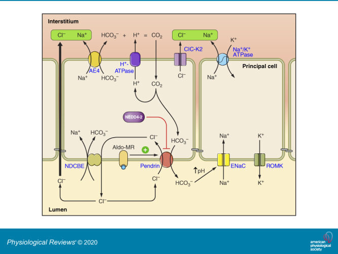
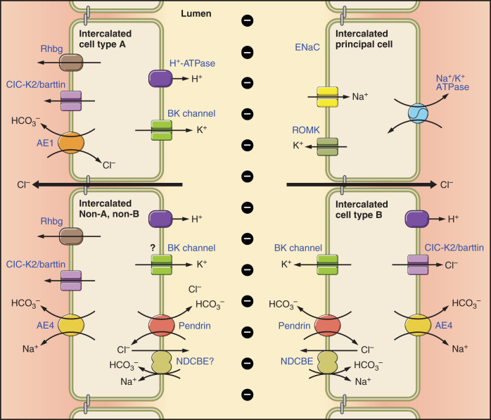
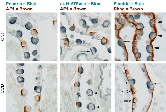
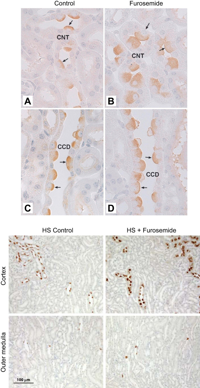
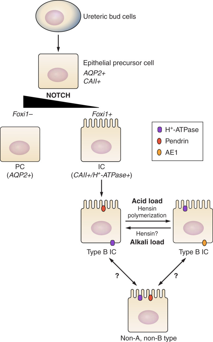
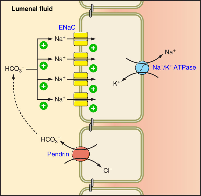
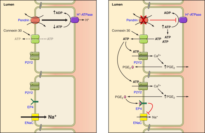
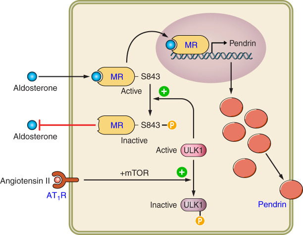
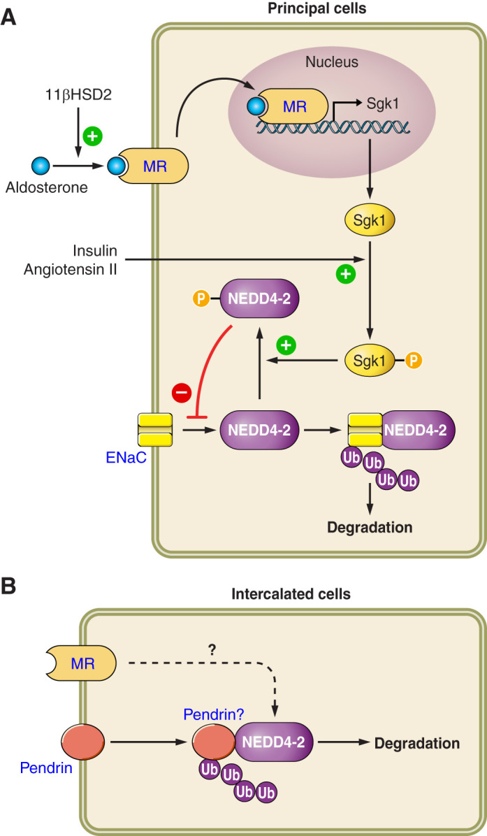
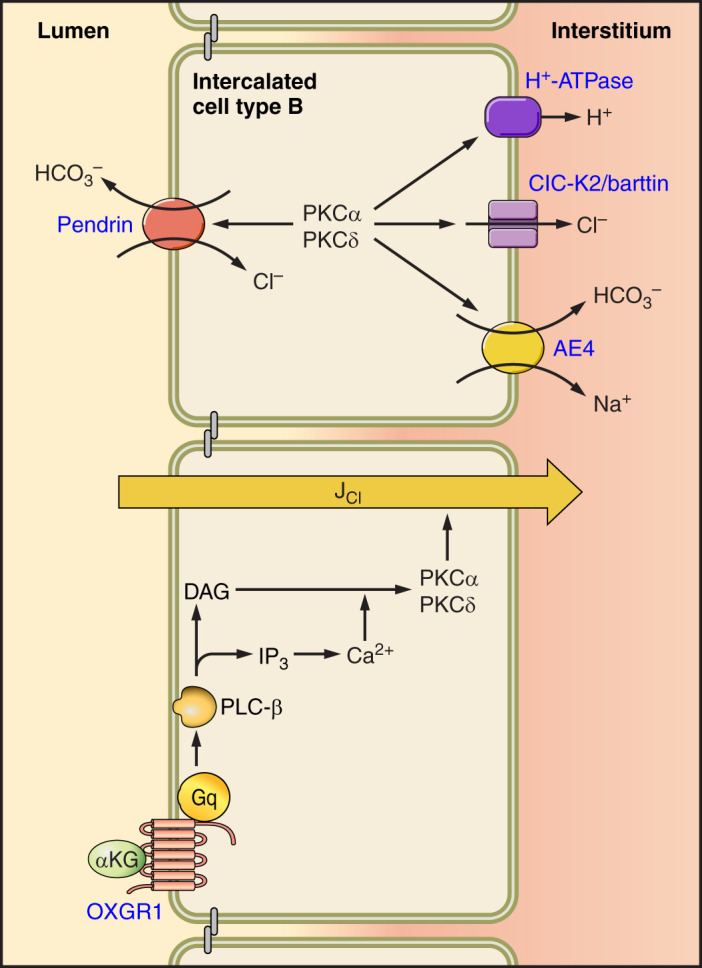
Intercalated cells (ICs) are found in the connecting tubule and the collecting duct. Of the three IC subtypes identified, type B intercalated cells are one of the best characterized and known to mediate Cl- absorption and HCO3- secretion, largely through the anion exchanger pendrin. This exchanger is thought to act in tandem with the Na+-dependent Cl-/HCO3- exchanger, NDCBE, to mediate net NaCl absorption. Pendrin is stimulated by angiotensin II and aldosterone administration via the angiotensin type 1a and the mineralocorticoid receptors, respectively. It is also stimulated in models of metabolic alkalosis, such as with NaHCO3 administration. In some rodent models, pendrin-mediated HCO3- secretion modulates acid-base balance. However, of probably more physiological or clinical significance is the role of these pendrin-positive ICs in blood pressure regulation, which occurs, at least in part, through pendrin-mediated renal Cl- absorption, as well as their effect on the epithelial Na+ channel, ENaC. Aldosterone stimulates ENaC directly through principal cell mineralocorticoid hormone receptor (ligand) binding and also indirectly through its effect on pendrin expression and function. In so doing, pendrin contributes to the aldosterone pressor response. Pendrin may also modulate blood pressure in part through its action in the adrenal medulla, where it modulates the release of catecholamines, or through an indirect effect on vascular contractile force. In addition to its role in Na+ and Cl- balance, pendrin affects the balance of other ions, such as K+ and I-. This review describes how aldosterone and angiotensin II-induced signaling regulate pendrin and the contribution of pendrin-positive ICs in the kidney to distal nephron function and blood pressure.

Keywords: Cl−/HCO3− exchange; ENaC; Slc26a4; blood pressure; intercalated cells; pendrin.


Conflict of interest statement

No conflicts of interest, financial or otherwise, are declared by the authors.

Figures

None
Graphical abstract
FIGURE 1.
FIGURE 1.
Cell types and transporters in the cortical collecting duct (CCD). Intercalated and principal cell ion transporter distribution within of the CCD is shown. In type B intercalated cells (ICs), the Na+-dependent Cl/HCO3 exchanger, NDCBE, mediates Na+ and HCO3 absorption, whereas pendrin mediates HCO3 secretion and Cl absorption. Through the action of these 2 transporters, Cl and HCO3 are recycled across the apical membrane. Net H+ and Cl exit occur across the basolateral plasma membrane through the Cl channel, ClC-K2, and the H+-ATPase. Na+ exits the cell through the Na+-HCO3 cotransporter, AE4. Type A ICs secrete H+ through the apical plasma membrane H+-ATPase with net HCO3 efflux across the basolateral plasma membrane through the Cl/HCO3 exchanger, AE1. Type A ICs also express the ammonia channel, Rhbg, in the basolateral regions of the cell. Principal cells absorb Na+ through the epithelial Na+ channel, ENaC, which exits across the basolateral plasma membrane through the Na+-K+-ATPase. In so doing, it generates a lumen-negative transepithelial voltage, which provides the driving force for net secretion of K+. Most likely, Cl is also absorbed through paracellular transport driven by the lumen-negative transepithelial voltage generated by the epithelial Na+ channel, ENaC. Non-A, non-B ICs are rare in mouse CCD. However, these cells express pendrin and the H+-ATPase on the apical plasma membrane and express ClC-K2, AE4, and Rhbg in the basolateral regions of the cell. Whether these cells express NDCBE is unclear.
FIGURE 2.
FIGURE 2.
Intercalated cell marker labeling in mouse cortical collecting duct (CCD) and connecting tubule (CNT). Characteristic immunolabeling of the three distinct intercalated cell subtypes in the CNT (top panels) and CCD (bottom panels) are shown by differential interference contrast microscopy (DIC). Type A intercalated cells express the basolateral anion exchanger AE1, apical H+-ATPase, and the basolateral ammonia transporter Rhbg. Type B intercalated cells express the apical anion exchanger, pendrin, and basolateral H+-ATPase, but not AE1 or Rhbg. Non-A, non-B intercalated cells express apical pendrin, apical H+-ATPase, and basolateral Rhbg, but not AE1. Left column: double labeling for pendrin (blue) and AE1 (brown), the latter of which is definitive for type A intercalated cells; apical pendrin (blue) is present in type B and non-A, non-B intercalated cells. Pendrin labeling is exclusively in AE1-negative cells. Middle column: double labeling for AE1 (brown) and the a4 subunit of H+-ATPase (blue). Type A intercalated cells (AE1-positive) have apical H+-ATPase label. Type B intercalated cells have basolateral H+-ATPase label (arrows), as well as diffuse apical label, which correlates with cytoplasmic vesicle labeling shown by immunogold electron microscopy. Type B intercalated cells are uncommon in the CNT, but represent virtually all of the non-A intercalated cells in the CCD. In the CNT, the majority of non-A intercalated cells have apical H+-ATPase label, but no basolateral label. These are non-A, non-B intercalated cells. Right column: double labeling for pendrin (blue) and Rhbg (brown). Type B intercalated cells and non-A, non-B intercalated cells, both pendrin-positive, can be discriminated by basolateral Rhbg expression. Non-A, non-B intercalated cells, which express basolateral Rhbg (arrowheads), are the predominant pendrin-positive cell type in the CNT. Type B intercalated cells do not express detectable Rhbg (arrows) and comprise virtually all of the pendrin-positive cells in the CCD. Rhbg immunolabel is also present in type A intercalated cells (open arrows), CNT cells, and CCD principal cells. [From Verlander and Clapp (216), with permission from Elsevier.]
FIGURE 3.
FIGURE 3.
Effect of furosemide on pendrin abundance and distribution in the cortex and medulla. Top panel shows renal cortical sections labeled for pendrin from mice that received 7 days of a NaCl-replete diet or diet and furosemide. As shown, in both the cortical collecting duct (CCD) (C and D) and connecting tubule (CNT) (A and B), cells that label for pendrin are much larger with more pendrin label in each cell in sections taken from the furosemide-treated mice. [From Pech et al. (140).] Bottom panel shows pendrin label in sections from the same mice at lower magnification. As shown, pendrin label is much more prominent in the cortex of furosemide-treated than of vehicle-treated mice. In the medulla, few pendrin-positive cells are observed in mice from either group.
FIGURE 4.
FIGURE 4.
Ontogeny of intercalated cell (IC) differentiation. ICs and principal cells (PCs) are derived from a common immature precursor (epithelial precursor cell) that is governed by Notch and Foxi1 signaling. In the absence of Notch signaling, the transcription factor Foxi1 is suppressed. However, the “terminal” phenotype of the various IC subtypes may interconvert under certain experimental conditions.
FIGURE 5.
FIGURE 5.
Pendrin-mediated HCO3 secretion modulates epithelial Na+ channel (ENaC) abundance and function. Pendrin mediates the secretion of HCO3, which stimulates ENaC-mediated Na+ absorption as well as ENaC subunit abundance.
FIGURE 6.
FIGURE 6.
Pendrin gene ablation modulates luminal ATP concentration, which changes epithelial Na+ channel (ENaC) abundance and function. With pendrin gene ablation, H+-ATPase abundance falls in the type B intercalated cell, thereby increasing intercalated cell ATP content. Luminal ATP concentration then rises through enhanced connexin 30-mediated ATP secretion. Luminal ATP acts on apical membrane purinergic receptors to stimulate calcium release, which increases prostaglandin E2 production (PGE2). PGE2 acts through a receptor-mediated process to reduce ENaC abundance and function.
FIGURE 7.
FIGURE 7.
Mechanism of intercalated cell (IC) mineralocorticoid receptor (MR) activation. When the IC MR is phosphorylated at S843, aldosterone binding to the MR is inhibited. However, angiotensin II acts through the angiotensin type 1 receptor (AT1R) and mammalian target of rapamycin (mTOR) to phosphorylate, and thereby inactivate, ULK1, a kinase that mediates MR S843 phosphorylation. By inhibiting ULK1, S843 phosphorylation does not occur, which facilitates aldosterone binding to and thus activation of the IC MR. Upon aldosterone binding, the IC MR is translocated to the nucleus, which promotes transcription of IC transporters such as pendrin.
FIGURE 8.
FIGURE 8.
The regulation of intercalated cell (IC) and principal cell function by Nedd4-2. In principal cells, the ubiquitin ligase Nedd4-2 associates with epithelial Na+ channel (ENaC), which promotes the ubiquitylation and thus degradation of this Na+ channel. Nedd4-2 is activated through dephosphorylation, which enables it to associate with target proteins such as ENaC. Conversely, when phosphorylated, which occurs with the phosphoactivation of Sgk1 that follows insulin or angiotensin II application, Nedd4-2 cannot associate with these target proteins. When a cell expresses 11β-hydroxysteroid dehydrogenase type 2 (11βHSD2), aldosterone can bind to the mineralocorticoid receptor (MR), resulting in its translocation to the nucleus, which induces gene transcription. In ICs, Nedd4-2 regulates pendrin subcellular distribution. Whether this occurs through a direct association of Nedd4-2 and pendrin, and whether Nedd4-2 acts downstream of the MR, remains to be determined.
FIGURE 9.
FIGURE 9.
α-Ketoglutarate (αKG) signal transduction pathway in intercalated cells (ICs). αKG acts on the OXGR1 receptor, which stimulates protein kinase C (PKC)-α through a Ca2+-dependent mechanism and stimulates PKC-δ to increase pendrin-dependent Cl absorption. The role of other type B IC transporters, known to modulate apical anion exchange, in αKG-induced Cl absorption is unknown. DAG, diacylglycerol; IP3, inositol 1,4,5-trisphosphate; PLC, phospholipase C. [Modified from Lazo-Fernandez et al. (103).]

References

    1. Ackermann D, Gresko N, Carrel M, Loffing-Cueni D, Habermehl D, Gomez-Sanchez C, Rossier BC, Loffing J. In vivo nuclear translocation of mineralocorticoid and glucocorticoid receptors in rat kidney: differential effect of corticosteroids along the distal tubule. Am J Physiol Renal Physiol 299: F1473–F1485, 2010. doi:10.1152/ajprenal.00437.2010. - DOI - PubMed
    1. Al-Awqati Q, Gao XB. Differentiation of intercalated cells in the kidney. Physiology (Bethesda) 26: 266–272, 2011. doi:10.1152/physiol.00008.2011. - DOI - PubMed
    1. Almeida AJ, Burg MB. Sodium transport in the rabbit connecting tubule. Am J Physiol Renal Physiol 243: F330–F334, 1982. doi:10.1152/ajprenal.1982.243.4.F330. - DOI - PubMed
    1. Alper SL, Natale J, Gluck S, Lodish HF, Brown D. Subtypes of intercalated cells in rat kidney collecting duct defined by antibodies against erythroid band 3 and renal vacuolar H+-ATPase. Proc Natl Acad Sci USA 86: 5429–5433, 1989. doi:10.1073/pnas.86.14.5429. - DOI - PMC - PubMed
    1. Amlal H, Petrovic S, Xu J, Wang Z, Sun X, Barone S, Soleimani M. Deletion of the anion exchanger Slc26a4 (pendrin) decreases apical Cl−/HCO3 exchanger activity and impairs bicarbonate secretion in kidney collecting duct. Am J Physiol Cell Physiol 299: C33–C41, 2010. doi:10.1152/ajpcell.00033.2010. - DOI - PMC - PubMed

Publication types

MeSH terms

LinkOut - more resources