Comparative Study
. 2020 Apr 9;61(4):50.
doi: 10.1167/iovs.61.4.50.

Hyperreflective Stress Lines and Macular Holes

Affiliations
Comparative Study

Hyperreflective Stress Lines and Macular Holes

Jackson M Scharf et al. Invest Ophthalmol Vis Sci. .

Abstract

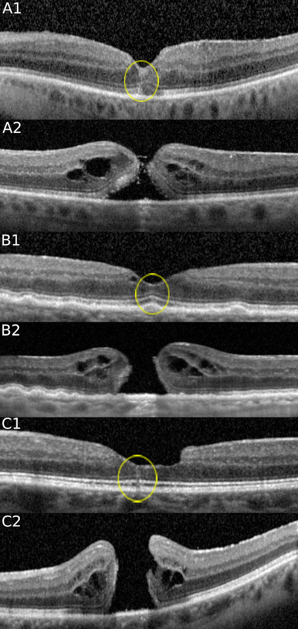
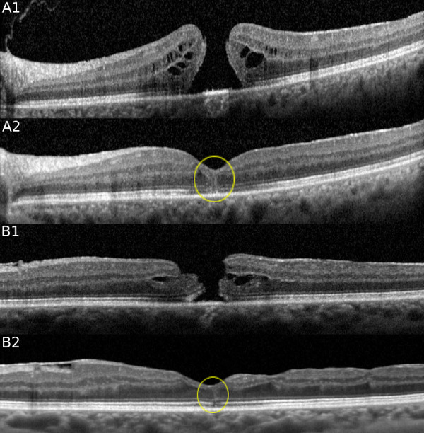
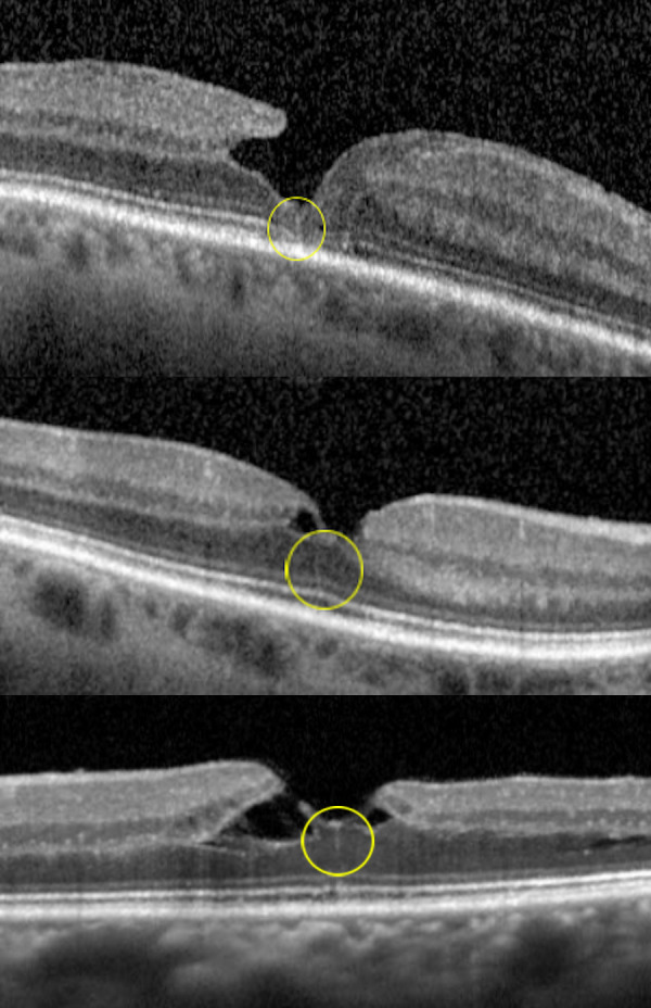
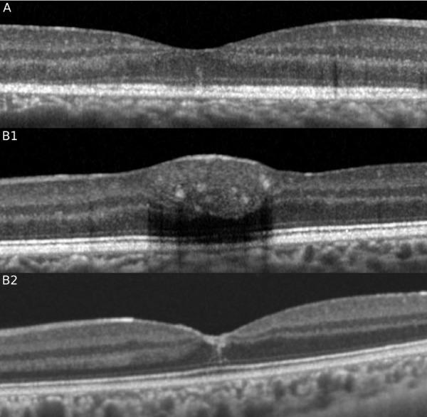
Purpose: To determine the prevalence of a central hyperreflective line in eyes with full-thickness macular holes (FTMH) and lamellar macular holes (LMH) and to elucidate the pathoanatomic importance of this optical coherence tomography (OCT) sign.

Methods: This retrospective analysis evaluated patients with FTMH and LMH at the Stein Eye Institute. Clinical data was collected and SD-OCT volume scans were analyzed for the presence of a central vertical hyperreflective line in 3 separate cohorts: patients with SD-OCT preceding FTMH development, patients with SD-OCT after pars plana vitrectomy (PPVT) treatment for FTMH, and patients with SD-OCT of LMH.

Results: In total, 93 eyes with FTMH and 88 eyes with LMH were identified. Of the 93 FTMH eyes, SD-OCT volume scans were available before development of the FTMH in 12 eyes. Of these, 6 (50%) displayed a vertical hyperreflective line preceding the development of the FTMH. Fifty-one eyes underwent PPVT with resolution of the FTMH, and 26 displayed a hyperreflective line after resolution (51%). Of the 88 eyes with LMH, 22 displayed a hyperreflective line (25%). All hyperreflective lines were noted at the central fovea.

Conclusions: SD-OCT illustrated the presence of a central vertical hyperreflective line preceding FTMH and after resolution of FTMH after PPVT in approximately one-half of cases, and concurrent with LMH in 25% of cases. This vertical hyperreflective line may represent an early SD-OCT marker for the development of FTMH, and may be a sign of central foveal dehiscence owing to disruption of the Muller cell cone.


Conflict of interest statement

Disclosure: J.M. Scharf, None; A. Hilely, None; R.C. Preti, None; C. Grondin, None; I. Chehaibou, None; G. Greaves, None; K. Tran, None; D. Wang, None; M.S. Ip, Alimera Sciences (C), Allergan (C), Boehringer Ingelheim (C), Genentech, Inc. (C), Omeros (C), Quark (C), ThromboGenics (C); J.P. Hubschman, Alcon (C), Allergan (C, S), Bausch & Lomb (C), Novartis (S); A. Gaudric, Novartis (C), ThromboGenics (C), Topcon (R); D. Sarraf, Amgen (C), Bayer (C), Genentech (C, F), Heidelberg (F), Novartis (S), Optovue (C, F), Regeneron (F), Topcon (F)

Figures

Figure 1.
Figure 1.
SD-OCT at the fovea of 3 cases before and after FTMH development. Note the presence of a hyperreflective line (yellow circle) preceding FTMH formation in each case. The hyperreflective vertical line extends from the area of the ellipsoid zone band to the ILM in the central fovea in each case. (Case A) In this 69-year-old man, a hyperreflective line at the fovea is noted in the left eye (A1) which progressed to a stage 2 FTMH (A2) just 2 months later. (Case B) In this 88-year-old man, a hyperreflective line at the fovea is noted in the right eye (B1), which evolved into a stage 4 FTMH (B2) 14 months later. (Case C) In this 62-year-old man, a hyperreflective line at the fovea is noted in the left eye (C1), which evolved into a stage 4 FTMH (C2) 4 months later.
Figure 2.
Figure 2.
SD-OCT of two cases of FTMH before and after PPVT. Note the presence of a hyperreflective line after resolution of the FTMH after PPVT. (Case A) A 63-year-old woman with a stage 4 FTMH (A1) in the left eye was treated with PPVT 3 weeks after the baseline OCT. A hyperreflective line at the fovea is noted with OCT at the 1-month follow-up (A2), which persisted for 9 months. (Case B) A 75-year old woman with a stage 4 FTMH (B1) in the left eye was treated with PPVT 2 weeks later. A hyperreflective line at the fovea (B2) is noted with OCT 8 months later.
Figure 3.
Figure 3.
SD-OCT illustrating the presence of a hyperreflective line (yellow circle) associated with a LMH. (Case A) (top) In this 68-year-old man with a lamellar hole in the right eye, a hyperreflective line is noted at the central fovea. (Case B) (middle) In this 61-year-old woman with a lamellar hole in the left eye, a thin hyperreflective line is noted at the central fovea. (Case C) (bottom) In this 71-year-old woman with a tractional lamellar hole and foveoschisis in the left eye, a hyperreflective line is noted at the central fovea.
Figure 4.
Figure 4.
SD-OCT showing the presence of the characteristic hyper reflective line in the central fovea in a case of multiple evanescent white dot syndrome associated with ellipsoid zone disruption (A) and in a separate case of macular hemorrhage (B1) after resolution (B2).

References

    1. Yamada E. Some structural features of the fovea centralis in the human retina. Arch Ophthalmol. 1969; 82: 151–159, doi:10.1001/archopht.1969.00990020153002. - DOI - PubMed
    1. Gass JDM. Müller cell cone, an overlooked part of the anatomy of the fovea centralis. Arch Ophthalmol. 1999; 117: 821, doi:10.1001/archopht.117.6.821. - DOI - PubMed
    1. Bringmann A, Pannicke T, Grosche J, et al. .. Müller cells in the healthy and diseased retina. Prog Retin Eye Res. 2006; 25: 397–424, doi:10.1016/j.preteyeres.2006.05.003. - DOI - PubMed
    1. Bringmann A, Syrbe S, Görner K, et al. .. The primate fovea: structure, function and development. Prog Retin Eye Res. 2018; 66: 49–84, doi:10.1016/j.preteyeres.2018.03.006. - DOI - PubMed
    1. Chung H, Byeon SH. New insights into the pathoanatomy of macular holes based on features of optical coherence tomography. Surv Ophthalmol. 2017; 62: 506–521, doi:10.1016/j.survophthal.2017.03.003. - DOI - PubMed

Publication types

MeSH terms