Review
. 2020 Jul 15;116(9):1600-1619.
doi: 10.1093/cvr/cvaa116.

Genetic basis and molecular biology of cardiac arrhythmias in cardiomyopathies

Affiliations
Review

Genetic basis and molecular biology of cardiac arrhythmias in cardiomyopathies

Ali J Marian et al. Cardiovasc Res. .

Abstract

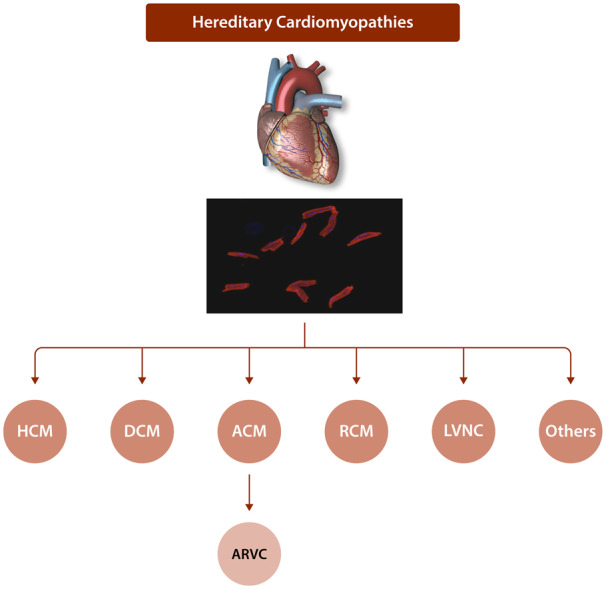
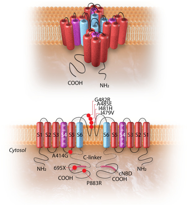
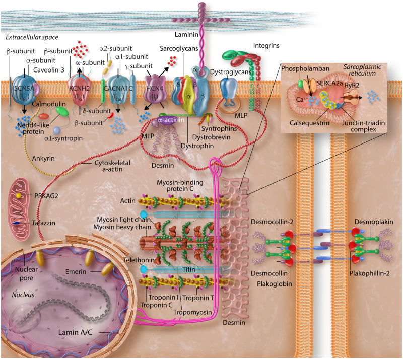
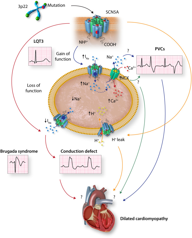
Cardiac arrhythmias are common, often the first, and sometimes the life-threatening manifestations of hereditary cardiomyopathies. Pathogenic variants in several genes known to cause hereditary cardiac arrhythmias have also been identified in the sporadic cases and small families with cardiomyopathies. These findings suggest a shared genetic aetiology of a subset of hereditary cardiomyopathies and cardiac arrhythmias. The concept of a shared genetic aetiology is in accord with the complex and exquisite interplays that exist between the ion currents and cardiac mechanical function. However, neither the causal role of cardiac arrhythmias genes in cardiomyopathies is well established nor the causal role of cardiomyopathy genes in arrhythmias. On the contrary, secondary changes in ion currents, such as post-translational modifications, are common and contributors to the pathogenesis of arrhythmias in cardiomyopathies through altering biophysical and functional properties of the ion channels. Moreover, structural changes, such as cardiac hypertrophy, dilatation, and fibrosis provide a pro-arrhythmic substrate in hereditary cardiomyopathies. Genetic basis and molecular biology of cardiac arrhythmias in hereditary cardiomyopathies are discussed.

Keywords: Genetics • Cardiomyopathy • Arrhythmias • Ion channels • Electrophysiology • Sudden death.


Figures

Figure 1
Figure 1
Classification of hereditary cardiomyopathies. Major forms for hereditary cardiomyopathies, which are classified based on their morphological and physiological features, are depicted. Adult cardiac myocytes are shown in the background to indicate that hereditary cardiomyopathies are primary disorders of cardiac myocytes.
Figure 2
Figure 2
The HCN4 channel and the topology of the HCN4 mutations associated with left ventricular non-compaction cardiomyopathy. The upper panel shows the 3D conformational structure of the HCN4 channel. The lower panel shows the liner structure of the HCN4 channel (two out of four subunits are shown). Each alpha-subunit is composed of six transmembrane helixes (S1–S6), a pore-forming loop, and intracellular N- and C-termini. The positively charged S4 helix (shown in purple) is the voltage sensor of the channel. The four S6 segments (shown in blue) of four-channel monomers together form the ion-conducting passage of the HCN4 channel. The C-terminus comprises of the C-linker (dotted line) and the cyclic nucleotide-binding domain (cNBD), which is connected to the channel core and mediates cyclic AMP-dependent changes in HCN channel gating. cAMP binding causes a conformation change that leads to the assembly of an active tetramer and channel opening. Red dots on the alpha-subunit shown on left indicate the sites of mutation associated with left ventricular non-compaction. The dot with a black dash indicates a truncation resulting from an insertion mutation leading to premature truncation of the protein at amino acid 695.
Figure 3
Figure 3
Schematic representation of the structural elements of cardiomyocytes implicated in the pathogenesis of arrhythmias in inherited cardiomyopathies. Protein constituents of cardiac myocyte structural proteins and ion channels are depicted to reflect the complexity of the interactions that mediate maintenance of normal cyclic excitation and contraction cycles throughout life.
Figure 4
Figure 4
Graphic illustration of putative mechanisms underlying dilated cardiomyopathy associated with SCN5A mutations. Gain-of-function (GoF) SCN5A variants are known to cause long QT3 syndrome. GoF variants could also increase INa current and to frequent premature ventricular contractions or ventricular tachycardia, which might contribute to left ventricular dilatation and dysfunction. Likewise, GoF variants could induce compensatory activation of the Na+/Ca2+ exchange protein, resulting in intracellular Ca2+ overload and consequently impaired excitation–contraction coupling and contractile dysfunction. Loss-of-function SCN5A variants are known to cause Brugada syndrome and cardiac conduction defects, the latter could lead to cardiac structural remodelling. SCN5A pathogenic variants could also result in proton leak into the cytosol, acidify the cytosol, and myocardial dysfunction. Experimental data to support these hypotheses are scant. INa, inward depolarizing sodium current; NaV1.5, voltage-gated sodium channel α-subunit; NCX, Na+/Ca2+ exchanger; PVC, premature ventricular contractions.

References

    1. McKenna W, Maron J, Barry J, Thiene G.. Classification, epidemiology, and global burden of cardiomyopathies. Circ Res 2017;121:722–730. - PubMed
    1. Marian AJ, Braunwald E.. Hypertrophic cardiomyopathy: genetics, pathogenesis, clinical manifestations, diagnosis, and therapy. Circ Res 2017;121:749–770. - PMC - PubMed
    1. Bagnall RD, Weintraub RG, Ingles J, Duflou J, Yeates L, Lam L, Davis AM, Thompson T, Connell V, Wallace J, Naylor C, Crawford J, Love DR, Hallam L, White J, Lawrence C, Lynch M, Morgan N, James P, Du Sart D, Puranik R, Langlois N, Vohra J, Winship I, Atherton J, McGaughran J, Skinner JR, Semsarian C.. A prospective study of sudden cardiac death among children and young adults. N Engl J Med 2016;374:2441–2452. - PubMed
    1. Maron BJ, Doerer JJ, Haas TS, Tierney DM, Mueller FO.. Sudden deaths in young competitive athletes: analysis of 1866 deaths in the United States, 1980. Circulation 2009;119:1085–1092. - PubMed
    1. O’Mahony C, Jichi F, Pavlou M, Monserrat L, Anastasakis A, Rapezzi C, Biagini E, Gimeno JR, Limongelli G, McKenna WJ, Omar RZ, Elliott PM, Hypertrophic Cardiomyopathy Outcomes Investigators. A novel clinical risk prediction model for sudden cardiac death in hypertrophic cardiomyopathy (HCM risk-SCD). Eur Heart J 2014;35:2010–2020. - PubMed

Publication types

MeSH terms

Substances