An official website of the United States government
The .gov means it’s official.
Federal government websites often end in .gov or .mil. Before
sharing sensitive information, make sure you’re on a federal
government site.
The site is secure.
The https:// ensures that you are connecting to the
official website and that any information you provide is encrypted
and transmitted securely.
1 Laboratory of Tumor Immunology and Biology, Center for Cancer Research, National Cancer Institute, National Institutes of Health, Bethesda, MD, USA.
2 Laboratory of Tumor Immunology and Biology, Center for Cancer Research, National Cancer Institute, National Institutes of Health, Bethesda, MD, USA. Electronic address: palenac@mail.nih.gov.
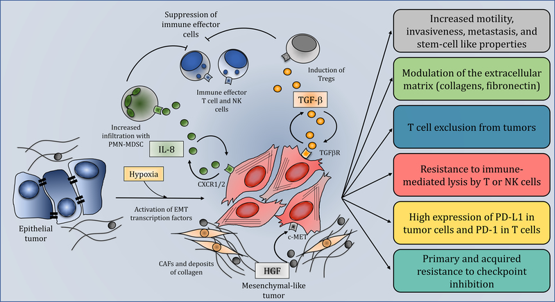
Tumor cell plasticity exhibited as an epithelial-mesenchymal transition (EMT) has been identified as a major obstacle for the effective treatment of many cancers. This process, which involves the dedifferentiation of epithelial tumor cells towards a motile, metastatic, and mesenchymal tumor phenotype, mediates resistance to conventional therapies and small-molecule targeted therapies. In this review, we highlight current research correlating the role of tumor plasticity with resistance to current immunotherapy approaches and discuss future and ongoing combination immunotherapy strategies to reduce tumor cell plasticity-driven resistance in cancer.
Figure 1.. Role of tumor cell plasticity in resistance to immunotherapy.
Epithelial tumor cells (blue…
Figure 1.. Role of tumor cell plasticity in resistance to immunotherapy.
Epithelial tumor cells (blue cells, left) acquire mesenchymal features through activation of EMT transcription factors or signaling initiated by soluble factors including IL-8 (circular green molecules) and TGF-β (circular orange molecules). Secreted IL-8 recruits PMN-MDSCs (green cells) to the tumor which in turn suppress immune effector T and NK cells (blue cells, top). TGF-β released by the tumor cells can induce Tregs (grey cells) which also contribute to the suppressive microenvironment. HGF (circular grey molecules) secreted by CAFs (yellow cells) signal through c-MET to promote tumor cell plasticity. Circular arrows indicate the ability of soluble factors to act in an autocrine fashion to maintain the mesenchymal phenotype of the tumor. The boxes on the right describe the various properties of mesenchymal tumor cells (red cells), including increased motility, invasiveness, and propensity to metastasize; ability to impact the tumor microenvironment (TME) via modulation of the extracellular matrix and promoting T cell exclusion from tumors; resistance to the cytotoxic activity of immune effector cells; upregulation of PD-L1. Altogether, these mechanisms synergize to promote primary and acquired resistance to immune checkpoint blockade.
Wood LD et al. (2007) The genomic landscapes of human breast and colorectal cancers. Science 318 (5853), 1108–13.
-
PubMed
Patel AP et al. (2014) Single-cell RNA-seq highlights intratumoral heterogeneity in primary glioblastoma. Science 344 (6190), 1396–401.
-
PMC
-
PubMed
Gerlinger M et al. (2012) Intratumor heterogeneity and branched evolution revealed by multiregion sequencing. N Engl J Med 366 (10), 883–892.
-
PMC
-
PubMed
Russo M et al. (2016) Tumor Heterogeneity and Lesion-Specific Response to Targeted Therapy in Colorectal Cancer. Cancer Discov 6 (2), 147–153.
-
PMC
-
PubMed
Diaz LA Jr. et al. (2012) The molecular evolution of acquired resistance to targeted EGFR blockade in colorectal cancers. Nature 486 (7404), 537–40.
-
PMC
-
PubMed