. 2020 Apr 27;21(9):3077.
doi: 10.3390/ijms21093077.

A Strategy for Personalized Treatment of iPS-Retinal Immune Rejections Assessed in Cynomolgus Monkey Models

Affiliations

A Strategy for Personalized Treatment of iPS-Retinal Immune Rejections Assessed in Cynomolgus Monkey Models

Shota Fujii et al. Int J Mol Sci. .

Abstract

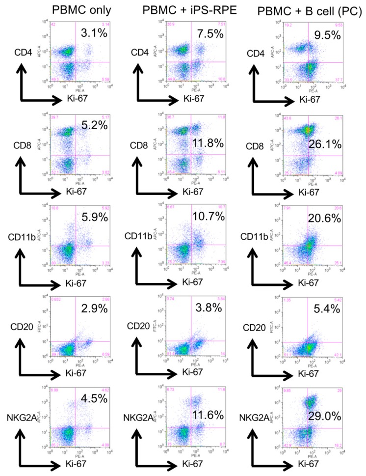
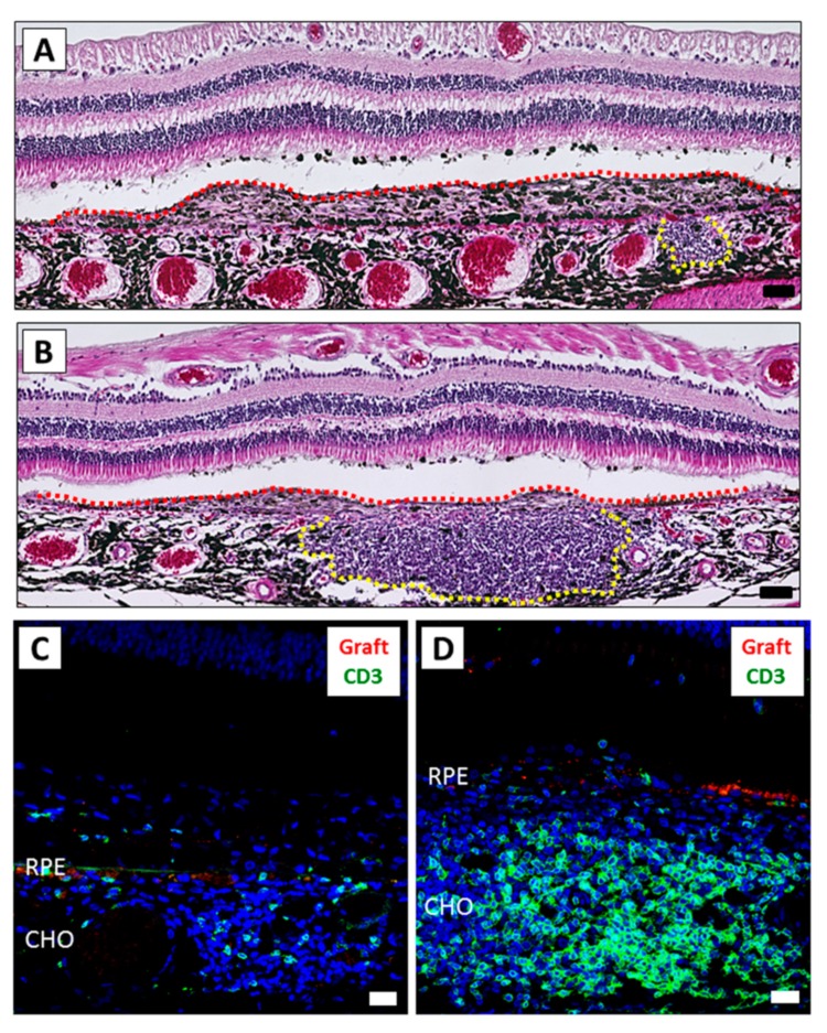
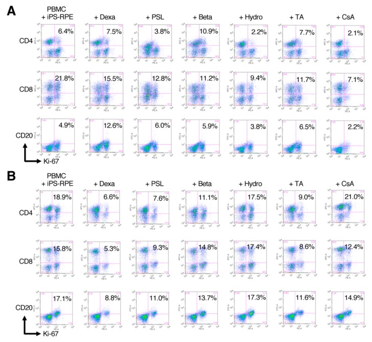
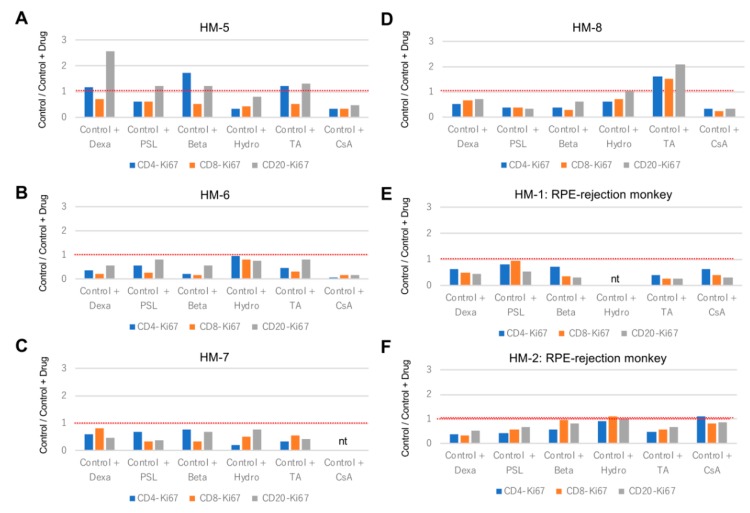
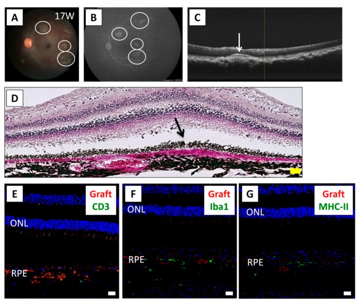
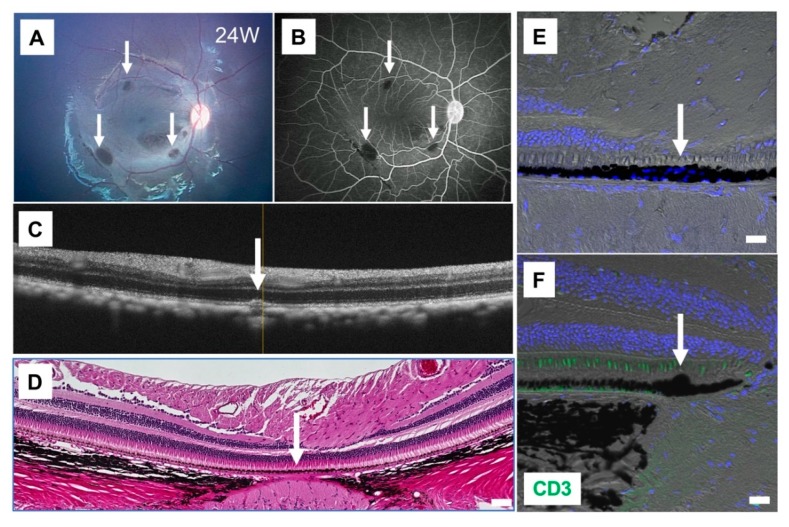
Recently, we successfully transplanted an autograft, or major histocompatibility complex (MHC)-matched allografts, from induced-pluripotent-stem-cell-derived retinal pigment epithelial (iPSC-RPE) cells in patients with age-related macular degeneration. However, there was an issue regarding immune rejection after transplantation. In this study, we established a preoperational in vitro "drug-lymphocytes-grafts immune reaction (Drug-LGIR)" test to determine the medication for immune rejection using host immunocompetent cells (lymphocytes) and transplant cells (target iPSC-RPE cells) together with different medications. The adequacy of the test was assessed by in vivo transplantation in monkey models together with medication based on in vitro data. In the results of Drug-LGIR tests, some drugs exhibited significant suppression of RPE cell-related allogeneic reactions, while other drugs did not, and the efficacy of each drug differed among the recipient monkeys. Based on the results of Drug-LGIR, we applied cyclosporine A or local steroid (triamcinolone) therapy to two monkeys, and successfully suppressed RPE-related immune rejections with RPE grafts, which survived without any signs of rejection under drug administration. We propose that our new preoperational in vitro Drug-LGIR test, which specifies the most efficacious medication for each recipient, is useful for controlling immune attacks with personalized treatment for each patient after retinal transplantation.

Keywords: drug; iPS cells; immune rejection; retinal pigment epithelial cells; transplantation.


Conflict of interest statement

The authors declare no competing interests.

Figures

Figure 1
Figure 1
Representative results of LGIR with peripheral blood mononuclear cells (PBMC) from a monkey. PBMCs (2 × 10⁶ cells/well) from a healthy monkey HM-1 were cultured with iPSC-RPE cells for 5 days. Before the assay, iPSC-RPE cells were irradiated (20 Gy) and 1 × 10⁴ cells were used for a 24-well culture plate. After 5 days of coculture, PBMC were harvested and stained with anti-CD4 (helper T cell-marker), anti-CD8 (cytotoxic T cell-marker), anti-CD11b (monocyte-, macrophage-, NK cell-, and granulocyte-marker), anti-CD20 (B cell-marker), anti-NKG2A (natural killer (NK) group 2 member A; NK cell-marker), and anti-Ki-67 (proliferation marker) antibodies. As a positive control (PC), irradiated allogenic B cells were used. The samples were analyzed by a fluorescence-activated cell sorting (FACS) flow cytometer. Numbers (%) in the scatterplots indicate double-positive cells (e.g., CD4/Ki-67).
Figure 2
Figure 2
The inflammatory difference between the right and left eyes after iPSC-RPE transplantation. The eyes of monkey HM-1 were histologically examined. (A,B) H&E staining of the right (A) and left (B) eye. Graft cells are indicated by the red dot-line. The mass of inflammatory cells is indicated by the yellow dot-line. The left eye exhibited severe inflammation compared to the right eye. Scale bars: 50 μm. (C,D) IHC of the right (C) and left (D) eye. A larger amount of CD3+ infiltration (green) was detected in the left eye compared to the right eye. PKH-positive live grafted RPE cells (red) were not detected. Scale bars: 20 μm. CHO: choroid.
Figure 3
Figure 3
Representative FACS results of Drug-LGIR in PBMC from healthy or transplanted monkeys. In the Drug-LGIR assay, PBMCs were cultured with human iPSC-RPE cells in the presence of indicated drugs for 5 days, and the suppressive effects of the drugs were evaluated by FACS of Ki-67 expressing cells. (A) The results of monkey HM-5 before transplantation. Hydrocortisone, prednisolone, and cyclosporine A exhibited significant suppression of the proliferation of the immune cell. By contrast, dexamethasone, betamethasone, and triamcinolone were not suppressive. (B) The results of monkey HM-2 that showed immune rejection against human iPSC-RPE cells in vivo. In the results of the in vitro assay, dexamethasone exhibited significant suppression of the immune reaction. Prednisolone and triamcinolone also exhibited suppressive effects. However, other drugs were not suppressive. Dexa: dexamethasone, PSL: prednisolone, Beta: betamethasone, Hydro: hydrocortisone, TA: triamcinolone, and CsA: cyclosporine A.
Figure 4
Figure 4
Summary of Drug-LGIR assay in monkeys. (AF) The results of Drug-LGIR assay with dexamethasone (Dexa), prednisolone (PSL), betamethasone (Beta), hydrocortisone (Hydro), triamcinolone (TA), and cyclosporine A (CsA) in PBMC from six monkeys: HM-5 (A), HM-6 (B), HM-7 (C), and HM-8 (D) that were not transplanted (n = 4) and HM-1 (E) and HM-2 (F) that showed RPE-rejection after transplantation (n = 2). The rate of proliferative immune cells indicated by expression of Ki-67 by FACS are shown in bar graphs to evaluate the suppressive effect of each drug. Red dotted-lines indicate the baseline (Control = 1.0, PBMC + iPS-RPE cells without drug). nt: not tested.
Figure 5
Figure 5
Results of iPSC-RPE transplantation while using local steroid therapy. (A) The fundus photograph of monkey HM-6 at 17 weeks after surgery, who was subjected to human iPSC-RPE transplantation with IVTA injection on day 0 and STTA injections at 4 and 12 weeks after transplantation. White circles show the transplanted sites. (B) Fluorescein angiography (FA) at late phase showed no hyperfluorescence around the grafts. No FA leakage was observed during the evaluation period. (C) Optical coherence tomography (OCT) revealed the presence of the cell aggregates of graft cells (arrow) in the subretinal space. (D) In H&E staining at 17 weeks after surgery, the graft cells (arrow) were observed in the subretinal space. Inflammatory cells were not obviously recognized. The overlying neurosensory retina was well preserved. Scale bar: 50 μm. (EG) IHC evaluation at 17 weeks after surgery. T cells were not observed in the retina (E). Although Iba1+ (F) and MHC class II+ cells (G) were observed around the grafts, little invasion of these antigen-presenting cells was observed throughout the retinal sections. PKH+ RPE cells (red), indicating live grafts, were detected in the subretinal space. Nuclei were stained by DAPI (blue). Scale bars: 20 μm. ONL: outer nuclear layer.
Figure 6
Figure 6
Results of iPSC-RPE transplantation while using cyclosporine A. (A) The fundus photograph of monkey HM-5 at 24 weeks after surgery, who was subjected to human iPSC-RPE transplantation with administration of cyclosporine A before the transplantation. Arrows show the transplanted sites. (B) Fluorescein angiography (FA) at late phase showed no hyperfluorescence around the grafts (arrows). No FA leakage was observed during the 24-week evaluation period. (C) Optical coherence tomography (OCT) showed the presence of cell aggregates of graft cells (arrow) in the subretinal space. (D) The graft cells (arrow) but not inflammatory cells were observed in the subretinal space by H&E staining. Scale bar: 50 μm. (EF) IHC with CD3 (green) and DAPI (blue). RPE grafts were detected in the subretinal space (arrows). CD3+ T cells were not observed in the retina (F). Scale bars: 20 μm.
Figure 7
Figure 7
Surgical complications seen after the iPSC-RPE transplantation: sterile endophthalmitis. (A) The fundus photograph of monkey HM-8 at 1 week after transplantation. Endophthalmitis was developed, which made the fundus invisible. (B) The inflammation disappeared around 3 weeks after surgery without any treatment. (C) H&E staining at 12 weeks after transplantation showed the graft (arrow) and epiretinal membrane (ERM; arrowhead). We diagnosed this case as triamcinolone-related sterile endophthalmitis. Scale bar: 100 μm.

References

    1. Okada A.A., Wakabayashi T., Morimura Y., Kawahara S., Kojima E., Asano Y., Hida T. Trans-Tenon’s retrobulbar triamcinolone infusion for the treatment of uveitis. Br. J. Ophthalmol. 2003;87:968–971. doi: 10.1136/bjo.87.8.968. - DOI - PMC - PubMed
    1. Mochizuki M. Immunotherapy for Behcet’s disease. Int. Rev. Immunol. 1997;14:49–66. doi: 10.3109/08830189709116844. - DOI - PubMed
    1. Ohno S., Nakamura S., Hori S., Shimakawa M., Kawashima H., Mochizuki M., Sugita S., Ueno S., Yoshizaki K., Inaba G. Efficacy, safety, and pharmacokinetics of multiple administration of infliximab in Behcet’s disease with refractory uveoretinitis. J. Rheumatol. 2004;31:1362–1368. - PubMed
    1. Diaz-Llopis M., Salom D., Garcia-de-Vicuna C., Cordero-Coma M., Ortega G., Ortego N., Suarez-de-Figueroa M., Rio-Pardo M.J., Fernandez-Cid C., Fonollosa A., et al. Treatment of refractory uveitis with adalimumab: A prospective multicenter study of 131 patients. Ophthalmology. 2012;119:1575–1581. doi: 10.1016/j.ophtha.2012.02.018. - DOI - PubMed
    1. Mandai M., Watanabe A., Kurimoto Y., Hirami Y., Morinaga C., Daimon T., Fujihara M., Akimaru H., Sakai N., Shibata Y., et al. Autologous Induced Stem-Cell-Derived Retinal Cells for Macular Degeneration. New Engl. J. Med. 2017;376:1038–1046. doi: 10.1056/NEJMoa1608368. - DOI - PubMed

MeSH terms