. 2020 Apr 27;11(5):475.
doi: 10.3390/genes11050475.

Telomere and Centromere Staining Followed by M-FISH Improves Diagnosis of Chromosomal Instability and Its Clinical Utility

Affiliations

Telomere and Centromere Staining Followed by M-FISH Improves Diagnosis of Chromosomal Instability and Its Clinical Utility

Radhia M'kacher et al. Genes (Basel). .

Abstract

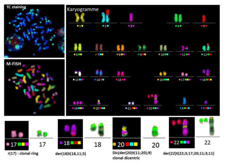
Dicentric chromosomes are a relevant marker of chromosomal instability. Their appearance is associated with telomere dysfunction, leading to cancer progression and a poor clinical outcome. Here, we present Telomere and Centromere staining followed by M-FISH (TC+M-FISH) for improved detection of telomere dysfunction and the identification of dicentric chromosomes in cancer patients and various genetic syndromes. Significant telomere length shortening and significantly higher frequencies of telomere loss and deletion were found in the peripheral lymphocytes of patients with cancer and genetic syndromes relative to similar age-matched healthy donors. We assessed our technique against conventional cytogenetics for the detection of dicentric chromosomes by subjecting metaphase preparations to both approaches. We identified dicentric chromosomes in 28/50 cancer patients and 21/44 genetic syndrome patients using our approach, but only 7/50 and 12/44, respectively, using standard cytogenetics. We ascribe this discrepancy to the identification of the unique configuration of dicentric chromosomes. We observed significantly higher frequencies of telomere loss and deletion in patients with dicentric chromosomes (p < 10-4). TC+M-FISH analysis is superior to classical cytogenetics for the detection of chromosomal instability. Our approach is a relatively simple but useful tool for documenting telomere dysfunction and chromosomal instability with the potential to become a standard additional diagnostic tool in medical genetics and the clinic.

Keywords: cancer; centromere; chromosomal instability; dicentric chromosome; genetic syndrome; telomere.


Conflict of interest statement

The authors declare no conflict of interest.

Figures

Figure 1
Figure 1
Cytogenetic detection of chromosomal aberrations. (A) R-banding, based on the morphological criteria of chromosomes, was the first and widely used technique for clinical cytogenetics. The detection of clonal aberration t (9;10)(q34;q2?6) in a renal-tract malformation patient without information on the precise breakpoint of chromosome 10 or the nature of translocation (balanced or unbalanced). (B) Telomere (red) and centromere (green) staining allows reliable classification of chromosomes and identification of chromosomal aberrations, such as t(9;10)(q34;q26.3), with precise localization of breakpoints confirming the reciprocal translocation. This particular reciprocal translocation involved the telomere region of chromosome 10. (C) The precise detection of the centromeric regions identifies all chromosomal aberrations, including dicentric chromosomes, in particular when both centromeres are very close, such as a tric (red arrow). The M-FISH technique does not stain the centromeric region. Telomere and Centromere staining followed by M-FISH technique (TC+M-FISH ) permits the reliable scoring and identification of all chromosomal aberrations, such as the dic (12;17), which could be mistaken for two chromosomes using only M-FISH. (D) Detection of a centric ring in circulating lymphocytes of a patient with a genetic syndrome using TC staining (c). This centric ring was undetectable by R-Banding(a) or M-FISH (b).
Figure 2
Figure 2
TC+M-FISH used to detect a complex karyotype in a case of acute lymphoblastic leukemia. This approach makes it possible to detect not only the translocation and complex exchange, but also the presence of a specific configuration of dicentric chromosomes with both the centromeres juxtaposed, such as dic(5;21)(p10;p10) and dic(21;22)(p10;p10) (arrow yellow and cyanine), or when the distance between both centromeres is very small (dic(10;17)(p10; p11).
Figure 3
Figure 3
Quantification of telomere length and telomere dysfunction. (A) Global quantification of telomere lengths in nuclei allows their assessment in a large number of cells and analysis of the intercellular heterogeneity of telomere lengths. An example of global quantification of telomere length in a healthy donor and a cancer patient is shown using TeloScore software. Our technique permits analysis of the mean telomere length, the distribution of fluorescence intensity in each quartile and the frequency of cells with telomeres < 5 kb. The distribution demonstrates the heterogeneity of telomere length. A significant difference between cells from the healthy donor and the cancer patient in the frequency of cells with drastic telomere shortening (< 5 kb) (black line) is demonstrated. However, mean telomere length (red line) is not always an appropriate indicator. (B) The use of cytogenetic slides for quantification of telomere length offers the possibility to detect telomere sequences in individual chromosomes, quantification of telomeres, and assessment of intrachromosomal variations of telomere length. Significant differences are observed in the intensity of each telomere in metaphases from a healthy donor and a cancer patient, the heterogeneity being greater for the cancer patient (C) The use of metaphases permits detection of telomere aberrations such as telomere loss, telomere deletion (the loss of two telomeres in the same arm), and the formation of telomere doublets. All these telomere aberrations are related to telomere dysfunction.
Figure 4
Figure 4
Telomere dysfunction in cancer patients and those with genetic syndromes. (A) Telomere length of healthy donors is age-dependent. The regression line indicates telomere shortening with age in healthy donors (79 pb per year; Y = 12.1−0.79X; R2 = 0.29). In cancer patients and those with genetic disorders, there is no significant correlation between telomere length and age. High individual variation is observed in telomere length of healthy donors, cancer patients, and genetic disorder patients. (B) Cancer patients and those with genetic syndromes show significantly shorter telomeres than healthy donors. (C) Analysis of the frequency of cells with short telomeres (<5 kb) reveals a significant difference between cancer patients and those with genetic disorders and healthy donors. (D) The frequency of telomere loss, the major telomere aberration that leads to telomere fusion and dicentric chromosome formation, is significantly higher in cancer patients and those with genetic syndromes than in healthy donors. (E) Similarly, the frequency of telomere deletions is significantly higher in cancer patients and those with genetic disorders than in healthy donors. (F) There is no significant difference in telomere doublet formation between healthy donors and patients with genetic disorders.
Figure 5
Figure 5
Variation of telomere dysfunction with age in healthy donors, cancer patients, and patients with genetic disorders: telomere dysfunction is relatively independent of age in all groups: (A) telomere loss, (B) telomere deletion, and (C) telomere doublet formation.
Figure 6
Figure 6
The presence of dicentric chromosome is associated with telomere dysfunction and complex karyotypes. Sequential analysis shows the presence of clonal dicentric chromosomes and centric rings in a mantle-cell lymphoma patient. These configurations are related to the presence of chromosomal aberrations related to breakage–fusion–bridge cycles, such as der(18) t (18,11;5) with an interstitial telomere of chromosome 18 and der(22) t (22;3;17;11;3;11).
Figure 7
Figure 7
Telomere dysfunction and dicentric chromosome formation. (A) High frequency of telomere loss and (B) deletion in patients with dicentric chromosomes compared to those without.
Figure 8
Figure 8
Overview of TC+M-FISH for the detection of chromosomal aberrations (A) Description of the TC+M-FISH approach. (B) Sensitivity of TC+M-FISH in the detection of dicentric chromosomes in cancer patients and those with genetic disorders compared to conventional cytogenetics. (C) Reporting time from the analysis of blood samples from patients, with or without complex karyotypes, using the TC+M-FISH approach compared to conventional cytogenetics (D) The cost (in euros) of the two approaches for the analysis of a simple and complex karyotype, based on the European situation.

References

    1. Burrell R.A., McGranahan N., Bartek J., Swanton C. The causes and consequences of genetic heterogeneity in cancer evolution. Nature. 2013;501:338–345. - PubMed
    1. Bakhoum S.F., Landau D.A. Chromosomal Instability as a Driver of Tumor Heterogeneity and Evolution. Cold Spring Harb. Perspect. Med. 2017;7:a029611. - PMC - PubMed
    1. Thompson S.L., Compton D.A. Chromosome missegregation in human cells arises through specific types of kinetochore-microtubule attachment errors. Proc. Natl. Acad. Sci. USA. 2011;108:17974–17978. - PMC - PubMed
    1. Pernot E., Hall J., Baatout S., Benotmane M.A., Blanchardon E., Bouffler S., El Saghire H., Gomolka M., Guertler A., Harms-Ringdahl M., et al. Ionizing radiation biomarkers for potential use in epidemiological studies. Mutat. Res. 2012;751:258–286. - PubMed
    1. McGranahan N., Burrell R.A., Endesfelder D., Novelli M.R., Swanton C. Cancer chromosomal instability: Therapeutic and diagnostic challenges. EMBO Rep. 2012;13:528–538. - PMC - PubMed