. 2020 Aug;14(8):1966-1981.
doi: 10.1038/s41396-020-0659-6. Epub 2020 Apr 29.

Diversity and dynamics of relevant nanoplanktonic diatoms in the Western English Channel

Affiliations

Diversity and dynamics of relevant nanoplanktonic diatoms in the Western English Channel

Laure Arsenieff et al. ISME J. 2020 Aug.

Abstract

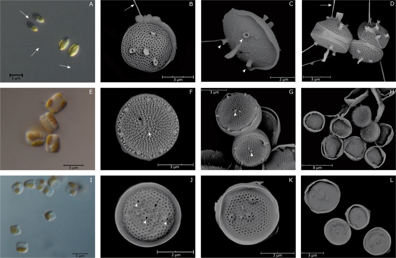
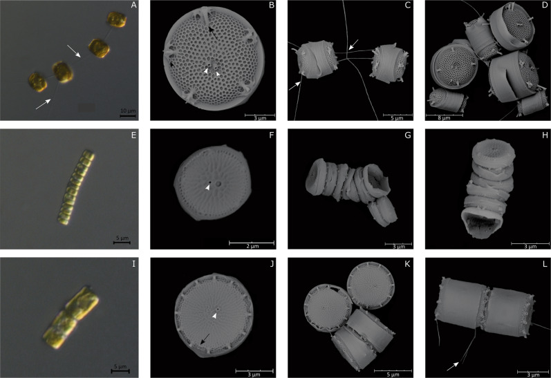
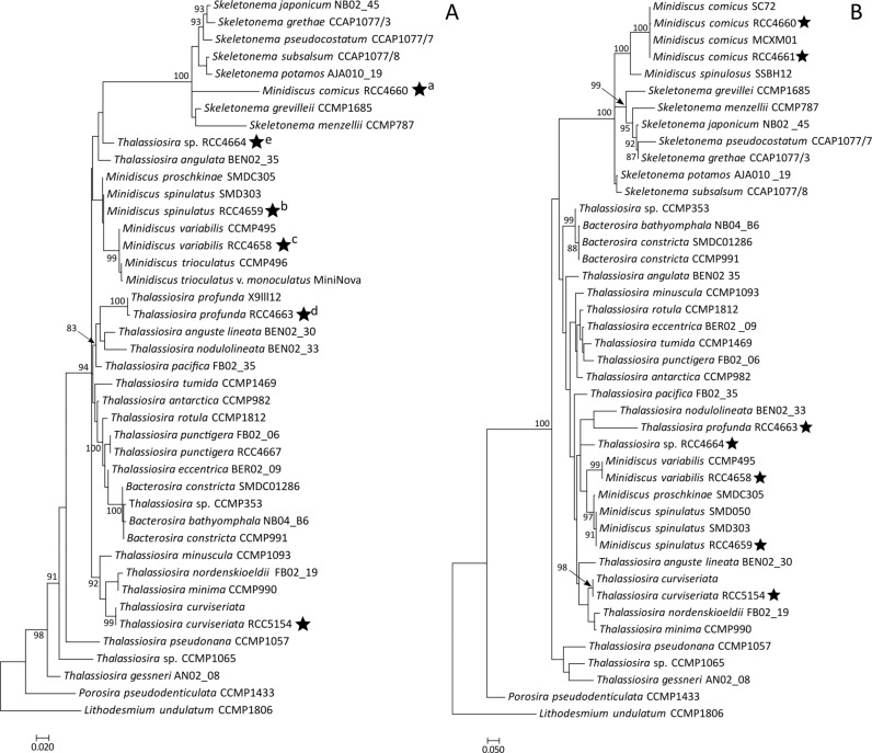
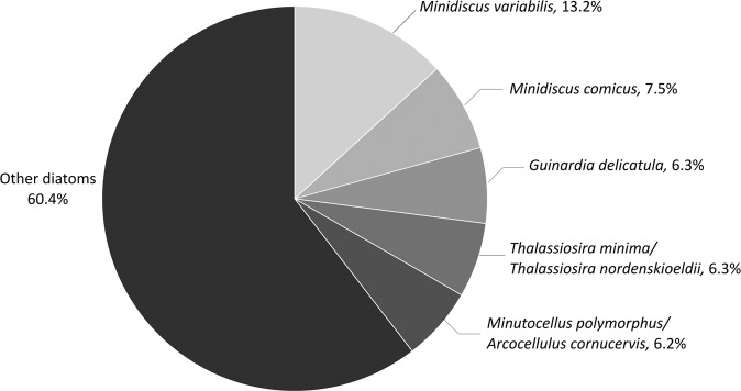
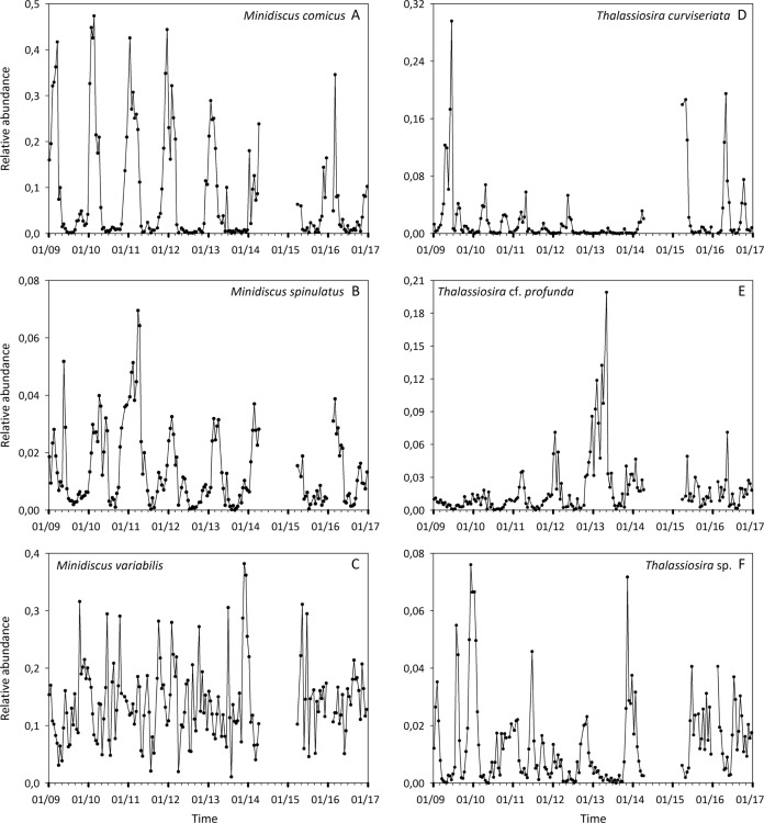
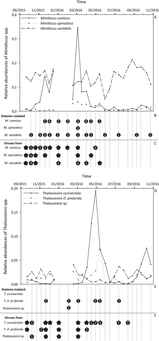
In the ocean, Bacillariophyta are one of the most successful protistan groups. Due to their considerable biogeochemical implications, diatom diversity, development, and seasonality have been at the center of research, specifically large-sized species. In comparison, nanoplanktonic diatoms are mostly disregarded from routine monitoring and are often underrepresented in genetic reference databases. Here, we identified and investigated the temporal dynamics of relevant nanodiatoms occurring in the Western English Channel (SOMLIT-Astan station). Coupling in situ and laboratory approaches, we revealed that nano-species from the genera Minidiscus and Thalassiosira are key components of the phytoplankton community that thrive in these coastal waters, but they display different seasonal patterns. Some species formed recurrent blooms whilst others were persistent year round. These results raise questions about their regulation in the natural environment. Over a full seasonal cycle at the monitoring station, we succeeded in isolating viruses which infect these minute diatoms, suggesting that these mortality agents may contribute to their control. Overall, our study points out the importance of considering nanodiatom communities within time-series surveys to further understand their role and fate in marine systems.


Conflict of interest statement

The authors declare that they have no conflict of interest.

Figures

Fig. 1
Fig. 1. LM and SEM micrographs of Minidiscus species.
M. comicus. a Pairs of cells connected by mucilaginous threads (LM). b, c, d External views of solitary cells (SEM). M. spinulatus. e Aggregated and solitary cells (LM). f, g, h External valve views. Note the Y-shaped ribs and the fultoportulae ring on the margin (SEM). M. variabilis. i Solitary cells (LM). j, k, l External view of valves. White arrows: threads connecting cells. Black arrows: Rimoportula. White arrowheads: Fultoportulae.
Fig. 2
Fig. 2. LM and SEM micrographs of Thalassiosira species.
T. curviseriata. a Chain of cells connected by threads (LM). b External valve view of solitary cells (SEM). Black arrowhead indicates the winged fultoportulae. c Girdle view of short chain of cells. d External view of large and small cells of T. curviseriata (SEM). T. cf. profunda. e Long chain of cells (LM). f Solitary cell in a valve view. Note the large areola adjacent to the central fultoportula (SEM). g, h Girdle views of chains (SEM). Thalassiosira sp. i Chain of cells (LM). j Valve external view of a solitary cell (SEM). k, l External views of solitary cells and of cells associated in pair (SEM). White arrows: threads connecting cells. Black arrows: Rimoportula. White arrowheads: Fultoportulae.
Fig. 3
Fig. 3. Phylogenetic position of dominant nanodiatoms isolated in the Western English Channel.
Phylogenetic rooted tree based on the 18S (a) and partial 28S (b) sequences of diatoms from the Thalassiosirales order. Porosira pseudodenticulata and Lithodesmium undulatum were taken as outgroups. The black stars indicate the positions of strains for which morphological characterizations were achieved in the frame of this study. Both Maximum Likelihood trees were generated using PhyML 3.0 with 1 000 replicates and a GTR + G + I substitution model according to the SMS analyses. Bootstrap values (%) greater than 80 are shown. Scale bars indicate the number of substitutions per site. Letters in superscript indicate that several strains had identical sequences. a: Minidiscus comicus strains RCC4660, RCC4661, RCC4662, and RCC5839 to RCC5859. b: Minidiscus spinulatus strains RCC4659, RCC5860 and RCC5861. c: Minidiscus variabilis strains RCC4657, RCC4658, RCC4665, RCC4666, and RCC5862 to RCC5880. d: Thalassiosira cf. profunda strains RCC4663 and RCC5881 to RCC5886. e: Thalassiosira sp. RCC4664 and RCC5887.
Fig. 4
Fig. 4. Relative contributions of the five most abundant OTUs related to Bacillariophyta at the SOMLIT-Astan station (2009–2016).
Taxonomic assignations were based on comparisons with the PR2 or NCBI databases and with the reference diatom strains described in this study.
Fig. 5
Fig. 5. Dynamics of the nanodiatoms isolated in the Western English Channel.
Variations in relative abundances of the OTUs related to a. Minidiscus comicus, b Minidiscus spinulatus, c Minidiscus variabilis, d Thalassiosira curviseriata, e Thalassiosira cf. profunda, and f Thalassiosira sp. at the SOMLIT-Astan station during the period 2009–2016. Note that metabarcoding data corresponding to 25 sampling dates (mainly between 2014 and 2015) were removed from the dataset (see “Material and methods”).
Fig. 6
Fig. 6. Seasonal dynamics of nanodiatoms and viruses isolated in the Western English Channel.
a, d Variations in the read relative abundances of the OTUs related to Minidiscus species (upper panel, a) and to Thalassiosira species (lower panel, d) at the SOMLIT-Astan station during the 2015–2016 period. b, e Respectively, Minidiscus and Thalassiosira isolates obtained during the period of our study. The number of isolated strains is indicated for each species and for each sampling date. c, f Virus isolates obtained respectively from Minidiscus and Thalassiosira cultures during the studied period. The success in the isolation procedure is indicated by pentagons while numbers indicate the number of viral strains still maintained in the laboratory (several strains were lost a few months after isolation). In b, c, e, and f, vertical dashed lines correspond to dates for which dilution series were carried out.
Fig. 7
Fig. 7. TEM micrographs of viruses and infected diatom.
a Negatively stained particles of the viral strain isolated in October 2015 on M. spinulatus RCC4659. As all virions displayed similar morphological features, micrographs of the other viral strains are not shown. b Ultrathin section of M. comicus infected by its associated virus. Arrowheads: viral particles accumulated in the host cytoplasm, F frustule, M mitochondrion, N nucleus, CH chloroplast.

References

    1. Nelson DM, Tréguer P, Brzezinski MA, Leynaert A, Quéguiner B. Production and dissolution of biogenic silica in the ocean: revised global estimates, comparison with regional data and relationship to biogenic sedimentation. Glob Biogeochem Cycles. 1995;9:359–72.
    1. Leblanc K, Arístegui J, Armand L, Assmy P, Beker B, Bode A, et al. A global diatom database—abundance, biovolume and biomass in the world ocean. Earth Syst Sci Data Discuss. 2012;4:147–85.
    1. Smetacek V. Diatoms and the ocean carbon cycle. Protist. 1999;150:25–32. - PubMed
    1. Tréguer P, Bowler C, Moriceau B, Dutkiewicz S, Gehlen M, Aumont O, et al. Influence of diatom diversity on the ocean biological carbon pump. Nat Geosci. 2018;11:27–37.
    1. Gómez F, Souissi S. Unusual diatoms linked to climatic events in the northeastern English Channel. J Sea Res. 2007;58:283–90.

Publication types