

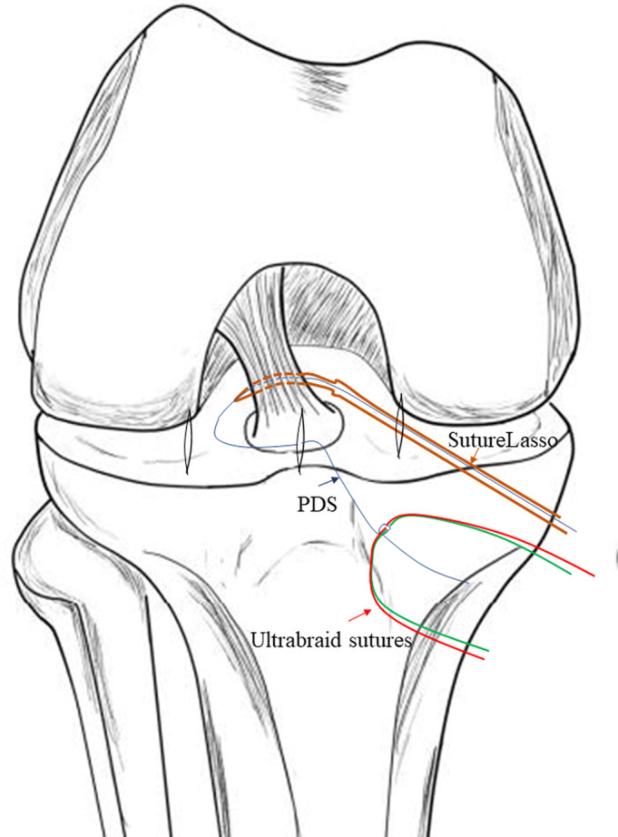
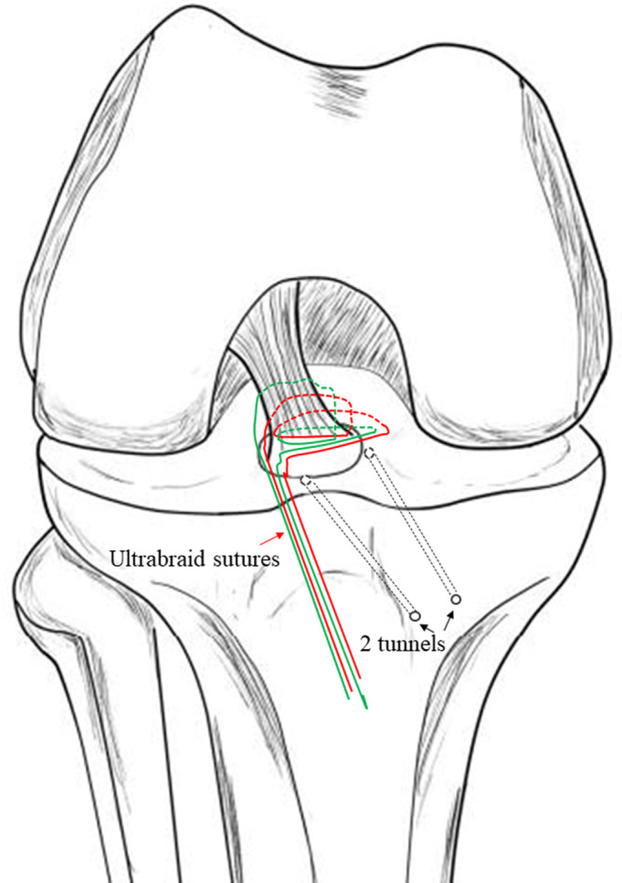
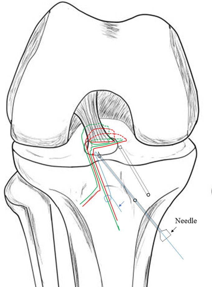
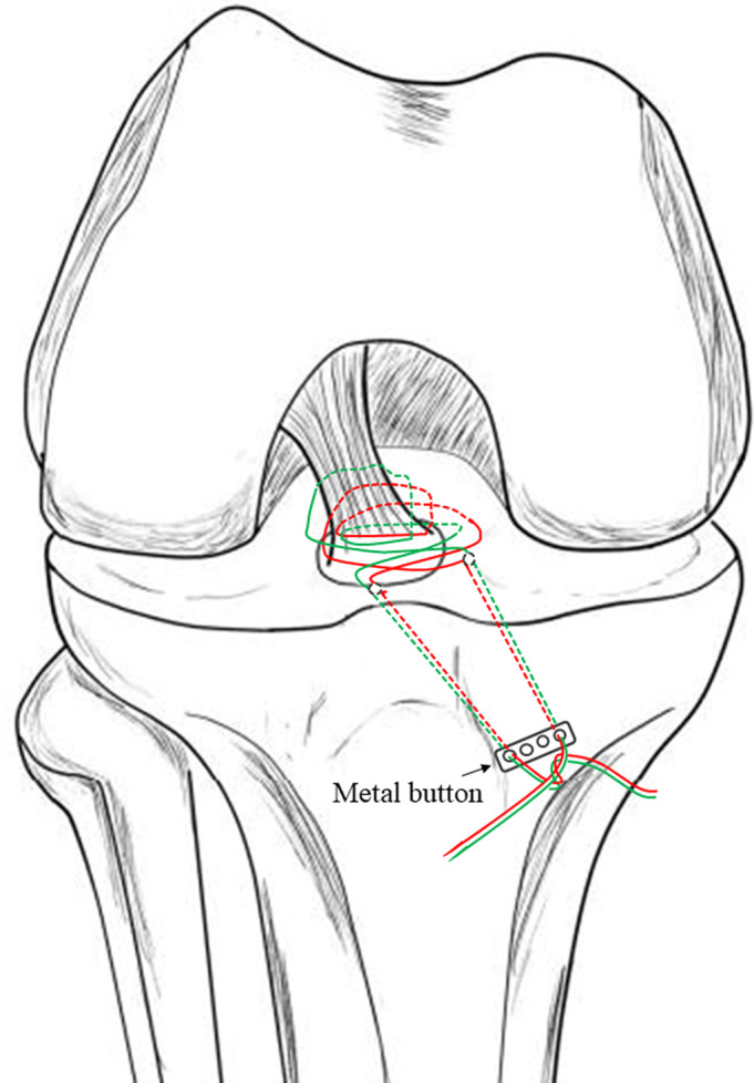
"Figure-of-Eight" Suture-Button Technique for Fixation of Displaced Anterior Cruciate Ligament Avulsion Fracture
- PMID: 32351046
- PMCID: PMC7307232
- DOI: 10.1111/os.12682
"Figure-of-Eight" Suture-Button Technique for Fixation of Displaced Anterior Cruciate Ligament Avulsion Fracture
Abstract
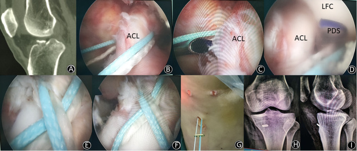
Objective: To assess the clinical results of the "figure-of-eight" suture-button technique in the arthroscopic treatment of anterior cruciate ligament (ACL) tibial avulsion fractures.
Methods: This was a retrospective study reviewing data from September 2013 to June 2019. A total of 27 patients (13 males and 14 females) who underwent arthroscopic "figure-of-eight" suture-button fixation for displaced ACL avulsion fractures were analyzed. The mean age of the patients in the sample was 15.8 years (10-29 years), with a mean follow-up of 24 months (6-48 months). According to Meyers-McKeever classification, 11 patients were classified as type III and 16 as type IV. All patients were evaluated following the guidelines of the radiological union, the Lysholm knee scoring scale, and the International Knee Documentation Committee (IKDC).
Results: Fractures were united within 3 months after surgery in all 27 cases. During the last follow-up, all the anterior drawer and Lachman tests were negative, except in 1 patient whose anterior drawer test was 1° positive. The range of motion was improved from 72.22° ± 27.92° before surgery to 137.78° ± 7.38° at the last follow-up (P < 0.05); the Lysholm score was improved from 45.81 ± 10.94 before surgery to 93.04 ± 5.66 at the last follow-up (P < 0.05); and the IKDC score was increased from 43.89 ± 11.16 before surgery to 90.26 ± 5.86 at the last follow-up (P < 0.05). In 1 patient, an inflammatory reaction was observed at the medial incision of the tibial tubercle; the symptoms disappeared with administration of antibiotics for 1 week. All patients returned to their preinjury physical activities at the last follow-up.
Conclusion: The "figure-of-eight" suture-button technique achieves a satisfactory clinical outcome and provides an effective method for the treatment of displaced ACL avulsion fractures.
Keywords: Anterior cruciate ligament; Arthroscopy; Avulsion; Fracture; Knee.
© 2020 The Authors. Orthopaedic Surgery published by Chinese Orthopaedic Association and John Wiley & Sons Australia, Ltd.
Figures
References
-
- Brunner S, Vavken P, Kilger R, et al Absorbable and non‐absorbable suture fixation results in similar outcomes for tibial eminence fractures in children and adolescents. Knee Surg Sports Traumatol Arthrosc, 2016, 24: 723–729. - PubMed
-
- Meyers MH, McKeever FM. Fracture of the intercondylar eminence of the tibia. J Bone Joint Surg Am, 1970, 52: 1677–1684. - PubMed
-
- Furlan D, Pogorelic Z, Biocic M, Juric I, Mestrovic J. Pediatric tibial eminence fractures: arthroscopic treatment using K‐wire. Scand J Surg, 2010, 99: 38–44. - PubMed
-
- Wiegand N, Naumov I, Vamhidy L, Not LG. Arthroscopic treatment of tibial spine fracture in children with a cannulated Herbert screw. Knee, 2014, 21: 481–485. - PubMed
-
- Parikh SN, Myer D, Eismann EA. Prevention of arthrofibrosis after arthroscopic screw fixation of tibial spine fracture in children and adolescents. Orthopedics, 2014, 37: e58–e65. - PubMed
MeSH terms
Grants and funding
LinkOut - more resources
Full Text Sources
Medical
