. 2020 Apr 21;26(15):1758-1774.
doi: 10.3748/wjg.v26.i15.1758.

PTEN-induced kinase 1-induced dynamin-related protein 1 Ser637 phosphorylation reduces mitochondrial fission and protects against intestinal ischemia reperfusion injury

Affiliations

PTEN-induced kinase 1-induced dynamin-related protein 1 Ser637 phosphorylation reduces mitochondrial fission and protects against intestinal ischemia reperfusion injury

Wasim Qasim et al. World J Gastroenterol. .

Abstract

Background: Intestinal ischemia reperfusion (I/R) occurs in various diseases, such as trauma and intestinal transplantation. Excessive reactive oxygen species (ROS) accumulation and subsequent apoptotic cell death in intestinal epithelia are important causes of I/R injury. PTEN-induced putative kinase 1 (PINK1) and phosphorylation of dynamin-related protein 1 (DRP1) are critical regulators of ROS and apoptosis. However, the correlation of PINK1 and DRP1 and their function in intestinal I/R injury have not been investigated. Thus, examining the PINK1/DRP1 pathway may help to identify a protective strategy and improve the patient prognosis.

Aim: To clarify the mechanism of the PINK1/DRP1 pathway in intestinal I/R injury.

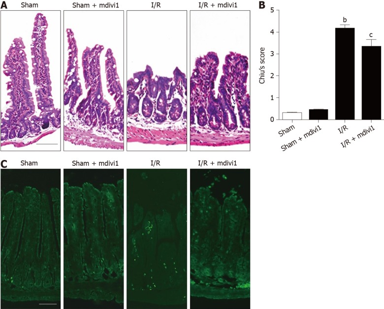
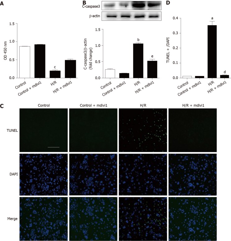
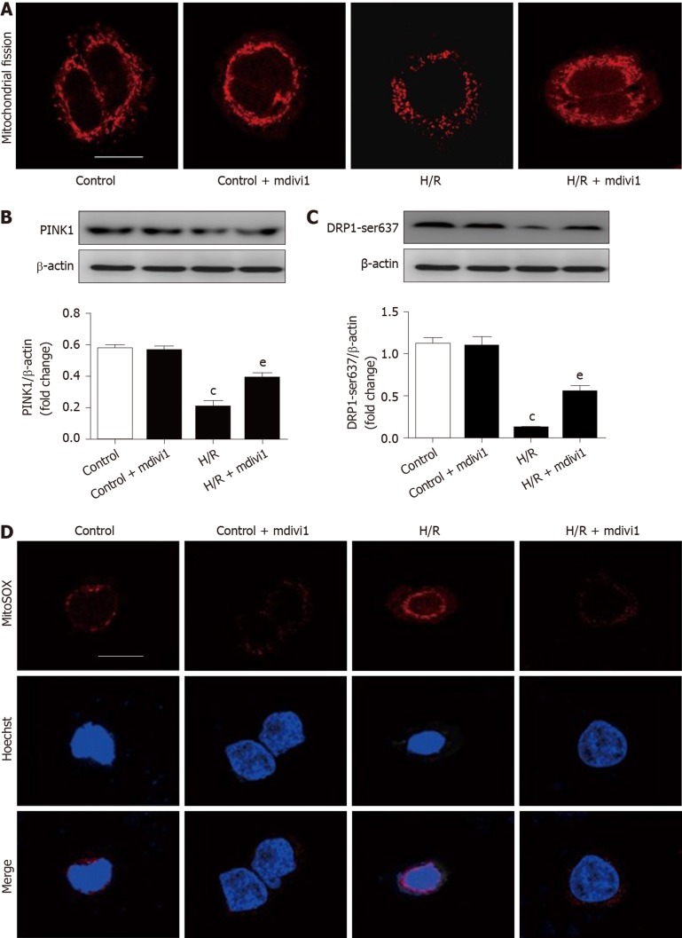
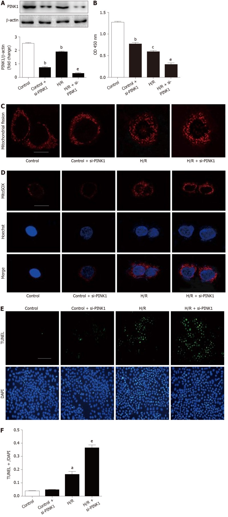
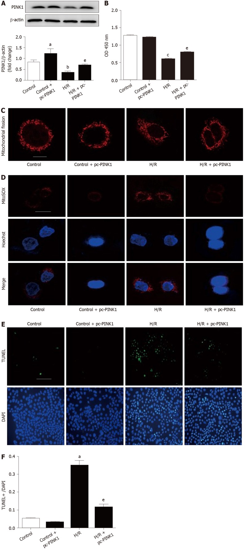
Methods: Male C57BL/6 mice were used to generate an intestinal I/R model via superior mesenteric artery occlusion followed by reperfusion. Chiu's score was used to evaluate intestinal mucosa damage. The mitochondrial fission inhibitor mdivi-1 was administered by intraperitoneal injection. Caco-2 cells were incubated in vitro in hypoxia/reoxygenation conditions. Small interfering RNAs and overexpression plasmids were transfected to regulate PINK1 expression. The protein expression levels of PINK1, DRP1, p-DRP1 and cleaved caspase 3 were measured by Western blotting. Cell viability was evaluated using a Cell Counting Kit-8 assay and cell apoptosis was analyzed by TUNEL staining. Mitochondrial fission and ROS were tested by MitoTracker and MitoSOX respectively.

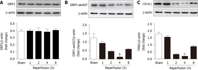
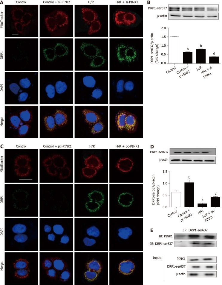
Results: Intestinal I/R and Caco-2 cell hypoxia/reoxygenation decreased the expression of PINK1 and p-DRP1 Ser637. Pretreatment with mdivi-1 inhibited mitochondrial fission, ROS generation, and apoptosis and ameliorated cell injury in intestinal I/R. Upon PINK1 knockdown or overexpression in vitro, we found that p-DRP1 Ser637 expression and DRP1 recruitment to the mitochondria were associated with PINK1. Furthermore, we verified the physical combination of PINK1 and p-DRP1 Ser637.

Conclusion: PINK1 is correlated with mitochondrial fission and apoptosis by regulating DRP1 phosphorylation in intestinal I/R. These results suggest that the PINK1/DRP1 pathway is involved in intestinal I/R injury, and provide a new approach for prevention and treatment.

Keywords: Apoptosis; Dynamin-related protein 1 ser637; Intestinal ischemia reperfusion injury; Mitochondrial fission; PTEN-induced putative kinase 1; Phosphorylation.


Conflict of interest statement

Conflict-of-interest statement: All the authors have nothing to disclose.

Figures

Figure 1
Figure 1
Expression of proteins in the intestine after various reperfusion times. A-C: Dynamin-related protein 1, dynamin-related protein 1 Ser637 and PTEN-induced kinase 1 expression at different reperfusion time points (n = 6). bP < 0.01, cP < 0.001 compared with the sham group. DRP1: Dynamin-related protein 1; PINK1: PTEN induced kinase 1.
Figure 2
Figure 2
Mdivi-1 treatment ameliorates tissue injury and apoptosis in intestinal ischemia/reperfusion. A: Hematoxylin and eosin staining of the small intestine (scale bar = 100 μm); B: Chui’s pathological score for the small intestine (n = 6); C: TUNEL staining of the small intestine (scale bar = 100 μm. bP < 0.01 compared with the sham group; cP < 0.05 compared with the ischemia/reperfusion group. I/R: Ischemia/reperfusion.
Figure 3
Figure 3
Mdivi-1 treatment protects cell viability and decreases apoptosis in Caco-2 hypoxia/reoxygenation cells. A: Cell viability was detected by the CCK8 method (n = 6); B: Representative Western blotting of cleaved caspase-3 protein expression in Caco-2 cells (n = 3); C: Effect of mdivi-1 on hypoxia/reoxygenation-induced cell apoptosis detected by TUNEL assay (scale bar = 100 μm); D: TUNEL positive cell ratio in hypoxia/reoxygenation-induced cell apoptosis (n = 3). aP < 0.05, bP < 0.01, cP < 0.001 compared with the control group; dP < 0.05, eP < 0.01 compared with the hypoxia/reoxygenation group. c-caspase 3: Cleaved caspase 3; H/R: Hypoxia/reoxygenation; TUNEL: Terminal-deoxynucleotidyl Transferase Mediated Nick End Labeling; DAPI: 4',6-diamidino-2-phenylindole.
Figure 4
Figure 4
Mdivi-1 administration inhibits mitochondrial fission and reactive oxygen species generation in Caco-2 hypoxia/reoxygenation cells. A: Deep Red FM in Caco-2 cells (scale bar = 10 μm); B, C: Dynamin-related protein 1 and dynamin-related protein 1 Ser637 expression upon mdivi-1 administration (n = 3); D: MitoSOX assay in Caco-2 cells (scale bar = 10 μm). cP < 0.001 compared with the control group; eP < 0.01 compared with the hypoxia/reoxygenation group. DRP1: Dynamin-related protein 1; PINK1: PTEN induced kinase 1; H/R: Hypoxia/reoxygenation.
Figure 5
Figure 5
Inhibition of PTEN-induced kinase 1 aggravates mitochondrial fission, reactive oxygen species accumulation and apoptosis in Caco-2 hypoxia/reoxygenation cells. A: The protein expression of PTEN- induced kinase 1 (PINK1) by Western blotting after PINK1 Small interfering RNA (siRNA) transfection (n = 3); B: Cell viability was detected by CCK-8 assay (n = 6); C: Deep Red FM in Caco-2 cells (scale bar = 10 μm); D: MitoSOX assay in Caco-2 cells (scale bar = 10 μm); E: PINK1 siRNA transfection increases intestinal cell apoptosis according to a TUNEL assay (length of the bar scale: 100 μm); F: PINK1 siRNA transfection increased intestinal cell apoptosis, as shown by the TUNEL positive cell ratio (n = 3). aP < 0.05, bP < 0.01, cP < 0.001 compared with the control group; eP < 0.01 compared with the H/R group. DRP1: Dynamin-related protein 1; PINK1: PTEN induced kinase 1; ROS: Reactive oxygen species; CCK-8: Cell Counting Kit-8; H/R: Hypoxia/reoxygenation; siRNA: Small interfering RNA; TUNEL: Terminal-deoxynucleoitidyl Transferase Mediated Nick End Labeling; DAPI: 4',6-diamidino-2-phenylindole.
Figure 6
Figure 6
Promotion of PTEN-induced kinase 1 expression decreases mitochondrial fission, reactive oxygen species accumulation and apoptosis in Caco-2 hypoxia/reoxygenation cells. A: Representative Western blotting of PTEN-induced kinase 1 (PINK1) protein expression in Caco-2 cells (n = 3); B: PINK1 overexpression increased Caco-2 cell viability according to CCK-8 assay (n = 6); C: PINK1 overexpression decreases mitochondrial fission during Hypoxia/reoxygenation (H/R) in Caco-2 cells according to MitoTracker staining (scale bars = 10 μm); D: PINK1 overexpression decreases reactive oxygen species levels detected by MitoSOX during H/R in Caco-2 cells (scale bars = 10 μm) (n = 3); E: TUNEL assay showing that PINK1 overexpression decreases apoptosis (scale bar = 100 μm); F: PINK1 overexpression decreases apoptosis, asindicated by the TUNEL-positive cell ratio (n = 3). aP < 0.05, bP < 0.01, cP < 0.001 compared with the control group; eP < 0.01 compared with the H/R group. PINK1: PTEN induced kinase 1; ROS: Reactive oxygen species; CCK-8: Cell Counting Kit-8; H/R: Hypoxia/reoxygenation; TUNEL: Terminal-deoxynucleoitidyl Transferase Mediated Nick End Labeling; DAPI: 4',6-diamidino-2-phenylindole.
Figure 7
Figure 7
PTEN-induced kinase 1 inhibits dynamin-related protein 1 recruitment on mitochondria by binding to Ser637. A, C: Mitochondrial localization of Dynamin-related protein 1 (DRP1) assessed under confocal a laser scanning microscope (green). Mitochondrial morphology was stained with MitoTracker Deep Red FM (red), and nuclei were stained with DAPI (blue) (scale bar = 10 μm); B, D: Representative Western blotting of DRP1 Ser637 protein expression in Caco-2 cells (n = 3); E: Coimmunoprecipitation assay of PTEN-induced kinase 1 with DRP1 Ser637. Representative immunoblot revealing the interaction of DRP1 Ser637 with PTEN-induced kinase 1. The input represents the total protein extracts used in immunoprecipitation. IB, immunoblotting; IP, immunoprecipitation, IgG, negative control. bP < 0.01 compared with the control group; dP < 0.01 compared with the H/R group. DRP1: Dynamin-related protein 1; PINK1: PTEN-induced kinase 1; H/R: hypoxia/reoxygenation; DAPI: 4',6-diamidino-2-phenylindole; IB: Immunoblot; IP: Immunoprecipitation.

References

    1. Acosta S. Epidemiology of mesenteric vascular disease: clinical implications. Semin Vasc Surg. 2010;23:4–8. - PubMed
    1. Mallick IH, Yang W, Winslet MC, Seifalian AM. Ischemia-reperfusion injury of the intestine and protective strategies against injury. Dig Dis Sci. 2004;49:1359–1377. - PubMed
    1. Stone JR, Wilkins LR. Acute mesenteric ischemia. Tech Vasc Interv Radiol. 2015;18:24–30. - PubMed
    1. Cheng J, Wei Z, Liu X, Li X, Yuan Z, Zheng J, Chen X, Xiao G, Li X. The role of intestinal mucosa injury induced by intra-abdominal hypertension in the development of abdominal compartment syndrome and multiple organ dysfunction syndrome. Crit Care. 2013;17:R283. - PMC - PubMed
    1. Li Y, Feng D, Wang Z, Zhao Y, Sun R, Tian D, Liu D, Zhang F, Ning S, Yao J, Tian X. Ischemia-induced ACSL4 activation contributes to ferroptosis-mediated tissue injury in intestinal ischemia/reperfusion. Cell Death Differ. 2019;26:2284–2299. - PMC - PubMed

MeSH terms