. 2020 Apr 15:11:532.
doi: 10.3389/fmicb.2020.00532. eCollection 2020.

The Biotherapeutic Potential of Lactobacillus reuteri Characterized Using a Target-Specific Selection Process

Affiliations

The Biotherapeutic Potential of Lactobacillus reuteri Characterized Using a Target-Specific Selection Process

Valeria Sagheddu et al. Front Microbiol. .

Abstract

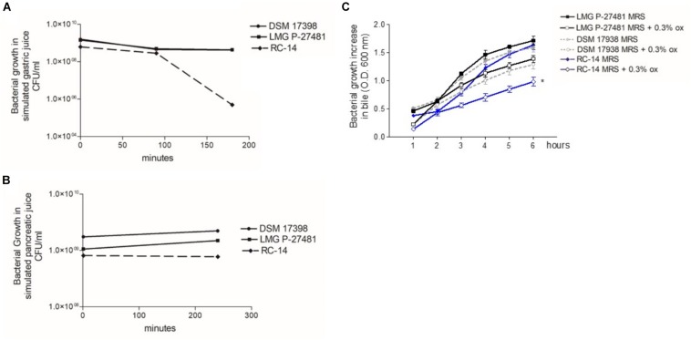
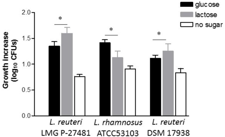
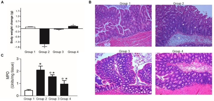
A growing body of clinical and experimental data supports the view that the efficacy of probiotics is strain-specific and restricted to particular pathological conditions, which means that newly isolated probiotic strains need to be targeted to a specific disease. Following national and international guidelines, we used a conventional in vitro experimental approach to characterize a novel strain of Lactobacillus reuteri, LMG P-27481, for safety (sensitivity to antibiotics and genome analysis) and putative efficacy (resistance to gastro-intestinal transit, adhesiveness, induction of cytokines, and release of antimicrobial metabolites). In vitro assays, which were carried out to examine the probiotic's effect on diarrhea (lactose utilization, inhibition of pathogens such as bacteria and Rotavirus), showed that it was more efficacious with respect to well-known reference strains in antagonizing Clostridioides difficile (CD). Data confirming that the probiotic can effectively treat CD colitis was gained from in vivo trials involving mice conditioned with large spectrum antibiotics before they were subjected to CD challenge. Two out of the three antibiotic-treated groups received daily LMG P-27481 for different time durations in order to simulate a preventive approach (LMG P-27481 administered prior to CD challenge) or an antagonistic one (LMG P-27481 administered after CD challenge). Both approaches significantly reduced, with respect to the untreated controls, CD DNA concentrations in caecum and C. difficile toxin titers in the gut lumen. In addition, LMG P-27481 supplementation significantly mitigated body weight loss and the extent of inflammatory infiltrate and tissue damage. The study results, which need to be confirmed by in vivo clinical trials, have demonstrated that the L. reuteri LMG P-27481 strain is a promising probiotic candidate for the treatment of CD infection.

Keywords: diarrhea; gut; intestine; lactobacilli; pathogen; probiotic.


Figures

FIGURE 1
FIGURE 1
The experimental protocol of L. reuteri LMG P-27481 treatment of C. difficile colitis in mice.
FIGURE 2
FIGURE 2
Lactobacillus reuteri LMG P-27481’s tolerance to in vitro gut transit gastric acid (A), pancreatic juice (B) and bile (C) exposure. (A) The probiotic’s growth, expressed in CFU/ml, following 0, 90, and 180 min of exposure to simulated gastric juice (n = 3 replicates for each strain). (B) The probiotic’s growth, expressed in CFU/ml, following 0 and 240 min of exposure to simulated pancreatic juice (n = 3 replicates for each strain). (C) Growth increase of probiotics, expressed as optical density OD 600 nm, following exposure for 6 h to bile salts. There are two curves for each strain tested: the reference growth standard on conventional laboratory medium MRS and the test curve reflecting the same medium supplemented with 0.3% vol/vol Oxgall.
FIGURE 3
FIGURE 3
Lactobacillus reuteri LMG P-27481 hydrogen peroxide release and adherence to human HT-29 monolayers. (A) Production of H2O2 by probiotics expressed as μg/ml of metabolite released by 109 viable CFUs after 24 h of incubation. (B) Antibiotic resistance image obtained by CARD showed the presence of nonperfect and strict and 111 loose hits. (C) Confluent HT-29 monolayers were washed and incubated with the probiotic strain (MOI 1:10) for 1 h at 37°C. To quantify adhering bacterial cells, the monolayers were extensively washed, lysed and aliquots seeded on MRS agar plates. Following 48 h of incubation in anaerobiosis, the colonies were enumerated and adhering bacteria expressed as the percentage of the total initial population of viable probiotic cells (n = 3 different experiments in duplicate). Data are presented as mean ± SD.
FIGURE 4
FIGURE 4
Lactobacillus reuteri LMG P-27481 induces an anti-inflammatory cytokine profile in human dendritic cells. The DCs obtained from differentiated peripheral blood monocytes were incubated (1:10 ratio) with the reported live bacterial strains for 1 h in medium without antibiotics. The cells were then collected, washed by centrifugation and incubated for an additional 23-h period in complete medium with antibiotics. Culture supernatants were collected. (A) IL-10 content quantified by ELISA. (B) IL-12p70 concentration assessed by ELISA. (C) Shows the anti-inflammatory index calculated as the ratio between IL-10 and IL-12p70 listed in Panels (A,B). Data are reported as mean ± SD on values obtained on 3 different donors. *P < 0.05 versus control and °P < 0.05 versus LMG P-27481.
FIGURE 5
FIGURE 5
Lactobacillus reuteri LMG P-27481 preferentially utilizes lactose as a carbon source. The effect of the presence of only glucose (black bars), only lactose (gray bars) or no sugars (open bars) as carbon sources, on growth performance of L. reuteri LMG P-27481, L. reuteri DSM 17938 or L. rhamnosus ATCC 53103, was calculated as growth increase in log10 CFUs. Bacteria were incubated for 18 h with 1% of each type of sugar and growth was recorded by decimal counts, (n = 3 replicates for each condition). Data are presented as mean ± SD. * indicates P < 0.05 versus growth in MRS containing glucose.
FIGURE 6
FIGURE 6
Lactobacillus reuteri LMG P-27481 inhibits pathogen growth in vitro. MRS broth was inoculated with 107 CFU/ml of the strain and incubated in anerobiosis for 24 h. MRS conditioned broths were sterile filtered, pH adjusted to 7 and inoculated at 37°C with either E. coli (107 CFU/ml) (A), Salmonella (107 CFU/ml) (B) or C. difficile (107 CFU/ml) (C). Aliquots of culture were collected after 6, 12 or 24 h and live bacteria quantified by seeding on proper Agar medium incubated at 37°C in aerobiosis for E. coli, Salmonella and anaerobiosis for C. difficile. Data are reported as mean ± SD on values obtained on three different determinations. *P < 0.05 versus not treated pathogen.
FIGURE 7
FIGURE 7
Lactobacillus reuteri LMG P-27481 reduces the severity of C. difficile colitis in a murine model. The mice were randomly divided in four experimental groups (Figure 1). Five days after intragastric (IG) challenge with C. difficile (105 CFU) the animals were sacrificed, and the colon content collected. (A) Body weight was measured at baseline (IG challenge) and every day for the following 5 days. Weight change was calculated as the variation in weight at sacrifice compared with that at the baseline. (B) Full thickness colonic specimens were fixed in 4% formalin, paraffin embedded and 8 μm thick sections stained with H&E. Tissue edema, inflammatory infiltrate and mucosal ulcers were evaluated and indicated with * in the Group 2 image. (C) Thickness colonic specimens were homogenized and used to quantify MPO activity as a marker of neutrophils infiltration. Data are reported as mean ± SD on values obtained on six animals for each experimental group. *P < 0.01 versus group 1 and °P < 0.05 versus group 2.
FIGURE 8
FIGURE 8
Lactobacillus reuteri LMG P-27481 reduces colonization in an antibiotic-induced C. difficile murine model. The mice were randomly divided into four experimental groups (Figure 1). Five days after intragastric (IG) challenge with C. difficile (105 CFU) the animals were sacrificed, and the caecum content collected. (A) Total DNA was extracted, and C. difficile specific DNA was quantified by qPCR. The data were quantified by the ΔΔCt method using 16S rDNA as the reference gene. (B) The caecal content was centrifuged, the clear supernatant sterile filtered and added to Vero cells monolayers to determine cytotoxicity. (C) Serial 1:2 dilutions of caecal content were added to Vero cells monolayers; the titer of C. difficile toxins was defined as the highest dilution causing >50% rounding 24 h after incubation. Data are reported as mean ± SD on values obtained on six animals for each experimental group. *P < 0.05 versus group 2.

References

    1. Abt M. C., McKenney P. T., Pamer E. G. (2016). Clostridium difficile colitis: pathogenesis and host defence. Nat. Rev. Microbiol. 14 609–620. 10.1038/nrmicro.2016.108 - DOI - PMC - PubMed
    1. Anderson D., McKay L. L. (1983). Simple and rapid method for isolating large plasmid DNA from lactic streptococci. Appl. Environ. Microbiol. 46 549–552. 10.1128/aem.46.3.549-552.1983 - DOI - PMC - PubMed
    1. Arnold J. W., Simpson J. B., Roach J., Bruno-Barcena J. M., Azcarate-Peril M. A. (2018). Prebiotics for lactose intolerance: variability in galacto-oligosaccharide utilization by intestinal Lactobacillus rhamnosus. Nutrients 10:1517. 10.3390/nu10101517 - DOI - PMC - PubMed
    1. Ausiello C. M., Cerquetti M., Fedele G., Spensieri F., Palazzo R., Nasso M., et al. (2006). Surface layer proteins from Clostridium difficile induce inflammatory and regulatory cytokines in human monocytes and dendritic cells. Microbes Infect. 8 2640–2646. 10.1016/j.micinf.2006.07.009 - DOI - PubMed
    1. Bath K., Roos S., Wall T., Jonsson H. (2005). The cell surface of Lactobacillus reuteri ATCC 55730 highlighted by identification of 126 extracellular proteins from the genome sequence. FEMS Microbiol. Lett. 253 75–82. 10.1016/j.femsle.2005.09.042 - DOI - PubMed

LinkOut - more resources